2,480
Views
16
CrossRef citations to date
0
Altmetric
Original Articles

The osteology of Magnosaurus nethercombensis (Dinosauria, Theropoda) from the Bajocian (Middle Jurassic) of the United Kingdom and a re-examination of the oldest records of tetanurans

Pages 131-146 | Received 23 Feb 2009, Accepted 05 Aug 2009, Published online: 12 Mar 2010

Figures & data

Figure 1 Megalosaurid dentaries. A-C, right dentary of Magnosaurus nethercombensis OUMNH J.12143/1b in medial (A, B) and dorsal (C) views; D, E, right dentary of Megalosaurus bucklandii OUMNH J.13505 in dorsal (D) and medial (E) views; F, right dentary of Duriavenator hesperis BMNH R332 in medial view; G, right dentary of Eustreptospondylus oxoniensis OUMNH J.13556 in medial view; H, left dentary of Dubreuillosaurus valesdunensis in medial view (reversed). In line drawing (B) light grey tone indicates tooth and dark grey tone indicates broken bone. Abbreviations: 3rd, third dentary alveolus; idp, interdental plate; mef, Meckelian fossa; meg, Meckelian groove; mfr, Meckelian foramen. Scale bars equal 100 mm. Parts of image are modified from CitationBenson et al. (2008) (D, E) and CitationBenson (2008a) (F).
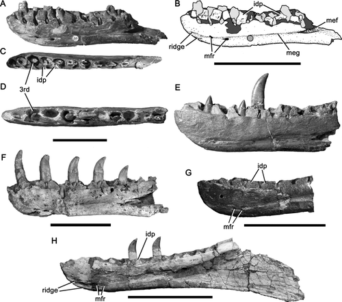
Figure 1 Megalosaurid dentaries. A-C, right dentary of Magnosaurus nethercombensis OUMNH J.12143/1b in medial (A, B) and dorsal (C) views; D, E, right dentary of Megalosaurus bucklandii OUMNH J.13505 in dorsal (D) and medial (E) views; F, right dentary of Duriavenator hesperis BMNH R332 in medial view; G, right dentary of Eustreptospondylus oxoniensis OUMNH J.13556 in medial view; H, left dentary of Dubreuillosaurus valesdunensis in medial view (reversed). In line drawing (B) light grey tone indicates tooth and dark grey tone indicates broken bone. Abbreviations: 3rd, third dentary alveolus; idp, interdental plate; mef, Meckelian fossa; meg, Meckelian groove; mfr, Meckelian foramen. Scale bars equal 100 mm. Parts of image are modified from CitationBenson et al. (2008) (D, E) and CitationBenson (2008a) (F).

Figure 2 Dentaries of Magnosaurus nethercombensis in lateral view. A, B, right dentary OUMNH J.12143/1b with magnified (x3) portions showing elongate foramina (A); C, left dentary OUMNH J.12143/1a. Abbreviations: for, foramen. Scale bar equals 100 mm.
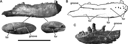
Figure 2 Dentaries of Magnosaurus nethercombensis in lateral view. A, B, right dentary OUMNH J.12143/1b with magnified (x3) portions showing elongate foramina (A); C, left dentary OUMNH J.12143/1a. Abbreviations: for, foramen. Scale bar equals 100 mm.

Table 1 Selected measurements (in millimetres) of Magnosaurus nethercombensis vertebrae. Abbreviations: anterior dv, dorsoventral height of anterior surface of centrum; anterior ml, mediolateral width of anterior surface of centrum; posterior dv, dorsoventral height of posterior surface of centrum; posterior ml, mediolateral width of posterior surface of centrum; i, measurement incomplete due to damage; e, measurement estimated from incomplete specimen.

Figure 3 Vertebrae of Magnosaurus nethercombensis. A-C, dorsal vertebra OUMNH J.12143/9 in right lateral (A), anterior (B), and ventral (C) views; D-F, proximal caudal vertebra OUMNH J.12143/8 in ventral (D), posterior (E), right lateral (F), and anterior (G) views. Abbreviations: chf, chevron facet. Scale bar equals 50 mm.

Figure 3 Vertebrae of Magnosaurus nethercombensis. A-C, dorsal vertebra OUMNH J.12143/9 in right lateral (A), anterior (B), and ventral (C) views; D-F, proximal caudal vertebra OUMNH J.12143/8 in ventral (D), posterior (E), right lateral (F), and anterior (G) views. Abbreviations: chf, chevron facet. Scale bar equals 50 mm.

Figure 4 Pelvic bones of Magnosaurus nethercombensis. A, B, partial right ilium OUMNH J.12143/10 in lateral (A) and medial (B) views; C, D, proximal portion of left pubis OUMNH J.12143/11 in lateral (C) and medial (D) views; E-G, shaft of right pubis OUMNH J.12143/3 in medial (E), anterior (F), and posterior (G) views. Abbreviations: ipr, ischial process; isp, ischial peduncle; pua, pubic apron. Scale bar equals 100 mm.
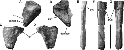
Figure 4 Pelvic bones of Magnosaurus nethercombensis. A, B, partial right ilium OUMNH J.12143/10 in lateral (A) and medial (B) views; C, D, proximal portion of left pubis OUMNH J.12143/11 in lateral (C) and medial (D) views; E-G, shaft of right pubis OUMNH J.12143/3 in medial (E), anterior (F), and posterior (G) views. Abbreviations: ipr, ischial process; isp, ischial peduncle; pua, pubic apron. Scale bar equals 100 mm.

Figure 5 Hindlimb bones of Magnosaurus nethercombensis. A-F and L, left femur OUMNH J.12143/7 in anterior (A, B), lateral (C), posterior (D, E), medial (F), and distal (L) views; G-K, right femur OUMNH J.12143/6 in anterior (G), medial (H), posterior (I-J), and distal (K) views; M-R, left tibia OUMNH J.12143/2 in anterior (M, N), lateral (O, P), proximal (Q), and distal (R) views. In line drawings (B, E, J, N, P) crossed hatching indicates matrix, light grey tone indicates plaster, and dark grey tone indicates broken bone. Abbreviations: cnc, cnemial crest; ctf, crista tibiofibularis; ffl, fibular flange; flx, flexor groove; lco, lateral condyle; mco, medial condyle; sab, suprastragalar buttress. Scale bar equals 200 mm.
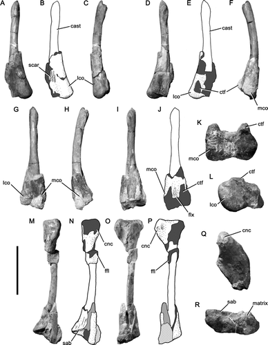
Figure 5 Hindlimb bones of Magnosaurus nethercombensis. A-F and L, left femur OUMNH J.12143/7 in anterior (A, B), lateral (C), posterior (D, E), medial (F), and distal (L) views; G-K, right femur OUMNH J.12143/6 in anterior (G), medial (H), posterior (I-J), and distal (K) views; M-R, left tibia OUMNH J.12143/2 in anterior (M, N), lateral (O, P), proximal (Q), and distal (R) views. In line drawings (B, E, J, N, P) crossed hatching indicates matrix, light grey tone indicates plaster, and dark grey tone indicates broken bone. Abbreviations: cnc, cnemial crest; ctf, crista tibiofibularis; ffl, fibular flange; flx, flexor groove; lco, lateral condyle; mco, medial condyle; sab, suprastragalar buttress. Scale bar equals 200 mm.

Figure 6 Theropod hindlimb bones from the Blue Lias of Charmouth, Dorset. A-F and L right femur BMNH 39496 in anterior (A, B), medial (C), posterior (D, E), lateral (F), and distal (L) views; G-K and M, right tibia BMNH 39496 in anterior (G), medial (H), posterior (I), lateral (J, K), and proximal (M) views; N-S, left femur GSM 109560 in medial (N, O), anterior (P, Q), posterior (R), and lateral (S) views. In line drawings (B, E, K, O, Q) dark grey tone indicates broken bone and light grey tone indicates reconstructed areas. Abbreviations: ail, anterior intermuscular line; cnc, cnemial crest; ctf, crista tibiofibularis; ffl, fibular flange; for, foramen; ft, fourth trochanter; ict, incisura tibialis; lco, lateral condyle; lt, lesser trochanter; mco, medial condyle; mdc, medial distal crest; trs, trochanteric shelf. Scale bars equal 100 mm.
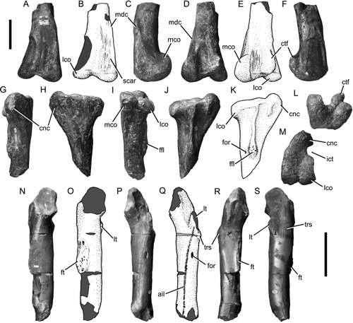
Figure 6 Theropod hindlimb bones from the Blue Lias of Charmouth, Dorset. A-F and L right femur BMNH 39496 in anterior (A, B), medial (C), posterior (D, E), lateral (F), and distal (L) views; G-K and M, right tibia BMNH 39496 in anterior (G), medial (H), posterior (I), lateral (J, K), and proximal (M) views; N-S, left femur GSM 109560 in medial (N, O), anterior (P, Q), posterior (R), and lateral (S) views. In line drawings (B, E, K, O, Q) dark grey tone indicates broken bone and light grey tone indicates reconstructed areas. Abbreviations: ail, anterior intermuscular line; cnc, cnemial crest; ctf, crista tibiofibularis; ffl, fibular flange; for, foramen; ft, fourth trochanter; ict, incisura tibialis; lco, lateral condyle; lt, lesser trochanter; mco, medial condyle; mdc, medial distal crest; trs, trochanteric shelf. Scale bars equal 100 mm.

Reprints and Corporate Permissions

Please note: Selecting permissions does not provide access to the full text of the article, please see our help page How do I view content?

To request a reprint or corporate permissions for this article, please click on the relevant link below:

Academic Permissions

Please note: Selecting permissions does not provide access to the full text of the article, please see our help page How do I view content?

Obtain permissions instantly via Rightslink by clicking on the button below:

If you are unable to obtain permissions via Rightslink, please complete and submit this Permissions form. For more information, please visit our Permissions help page.