Figures & data
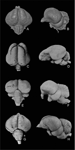
Figure 1 Endocranial casts of Eocene fossil birds in left lateral (top row) and dorsal (bottom row) views. A, B, Odontopteryx toliapica; C, D, Prophaethon shrubsolei; E, F, ‘Numenius’ gypsorum. A–D are virtual endocranial casts segmented from μCT scans. E and F are modified after CitationDechaseaux (1970).
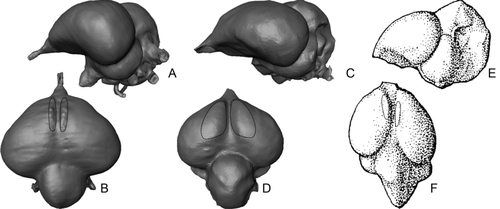
Figure 2 μCT visualization of the skull of Halcyornis toliapicus (NHMUK A130) rendered semi-transparent to reveal the virtual endocranial cast (blue) and inner ear (red). The skull is shown in ‘alert’ posture, based on the position of the horizontal semicircular canal (sensu CitationWitmer et al., 2003; CitationMilner & Walsh 2009).
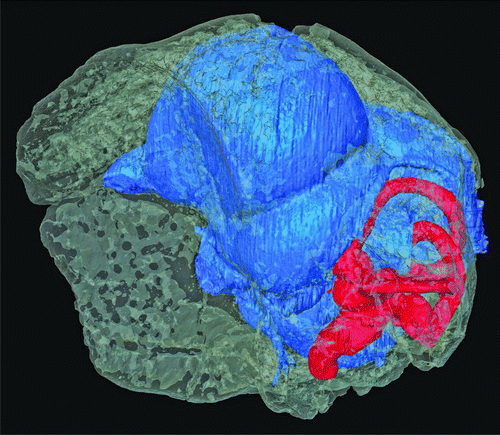
Table 1 Measurements taken from virtual endocast of Halcyornis toliapicus (NHMUK A130). All measurements and proportions are rounded to the nearest integer. Note that the brain region volumes total less than 100% because this method does not include space inside the endocranial cast beyond the limits of the external expression of the region. Similarly, the brain region surface area total is less than 100% because beyond the absolute external limits of some regions there are poorly differentiated areas (e.g. diencephalon) that were not included.
Figure 3 Virtual endocranial cast of Halcyornis toliapicus (NHMUK A130) in A, dorsal; B, ventral; C, caudal; D, rostral; E, left lateral views. See text for list of abbreviations. Scale bar = 5 mm.
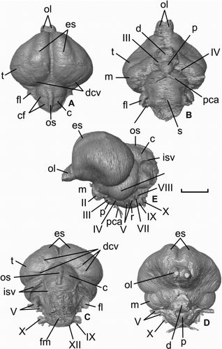
Figure 4 Virtual rendering of the left endosseous inner ear labyrinth of Halcyornis toliapicus (NHMUK A130) in: A, rostral; B, lateral; C, caudal; D, medial; E, dorsal views. See text for list of abbreviations. Scale bar = 5 mm.
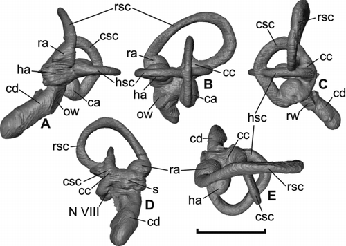
Figure 5 Hearing sensitivity of Halcyornis toliapicus (A, hearing range; B, mean hearing) based on regressions calculated from data derived from living bird and reptile species (see CitationWalsh et al. 2009; reproduced with permission of Royal Society Publishing).
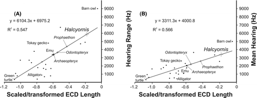
Table 2 Dimensions and intersections of the endosseous inner ear labyrinth of Halcyornis toliapicus.
Figure 6 Comparison of the virtual endocranial cast of Halcyornis toliapicus (NHMUK A130) with fresh brain material of taxa previously suggested to be closely related, in dorsal (left column) and left lateral (right column) views. A, B, Halcyornis toliapicus; C, D, Psittacus erithacus; E, F, Coracius garrulus; G, H, Larus argentatus. Note that the semicircular vein seen in A and B is an impression on the endocranium and is therefore not visible in D, F or H. C–H modified after CitationStingelin (1957).