Figures & data
Figure 1. Geographical distributions of Rhynchocephalia of the Upper Jurassic Morrison Formation discussed within the main text and Supplemental material, including type localities of the four named taxa. A, map of North America with the three US states having produced Late Jurassic rhynchocephalians shaded in grey; B, enlargement of Wyoming, Utah and Colorado with the approximate placement of the fossil localities as numbered diamonds. Solid black diamonds designate type localities; open diamonds represent other important localities discussed in the text. Localities numbered as follows: 1, Fox Mesa, Big Horn County (type locality of Opisthiamimus gregori gen. et sp. nov.); 2, Ninemile Hill, Carbon County; 3, Quarry 9, Como Bluff, Albany County (type locality of Opisthias rarus and Theretairus antiquus); 4, Rainbow Park and other nearby localities, Dinosaur National Monument, Uintah County; 5, Fruita Paleontological Area, Mesa County (type locality of Eilenodon robustus). C, north-central Wyoming, enlarged from B, highlighting the type locality of O. gregori gen. et sp. nov. (USNM loc. 42188, Fox Mesa; encircled star). Abbreviations: CDP, US census-designated place; CO, Colorado; loc., locality; USNM, National Museum of Natural History (formerly United States National Museum), Smithsonian Institution, Washington, DC, USA; UT, Utah; WY, Wyoming.
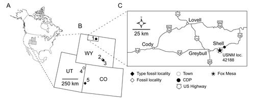
Figure 2. Anterior portion of the skull and lower jaws of the holotype specimen (USNM PAL 722041, ‘skull block’) of Opisthiamimus gregori gen. et sp. nov. A–C, extended depth of field stereophotopairs in A, dorsal, B, ventral and C, right lateral views. D–F, interpretive camera lucida drawings for A–C, respectively. Abbreviations: Co, coronoid; Den, dentary; den.can.t, dentary successional caniniform tooth; den.co.pr, coronoid process of the dentary; den.sym.s, symphyseal surface of the dentary; Ecpt, ectopterygoid; ?Ept, possible epipterygoid; Fr, frontal; Jug, jugal; Mx, maxilla; mx.can.t, successional caniniform tooth of the maxilla; Na, nasal; orb, orbit; Pal, palatine; pal.t, palatine tooth; Par, parietal; Pmx, premaxilla; Pofr, postfrontal; Porb, postorbital; Prfr, prefrontal; Pt, pterygoid; Sq, squamosal; Vo, vomer; vo.t, vomer tooth.
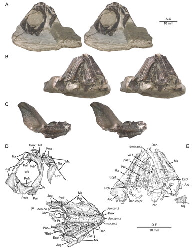
Figure 3. Posterior portion of the skull and lower jaws of the holotype specimen (USNM PAL 722041, ‘skeletal block’) of Opisthiamimus gregori gen. et sp. nov. A, extended depth of field (EDF) stereophotopair in dorsal view; B, interpretive camera lucida drawing for A; C, EDF stereophotopair in anterodorsal view. Abbreviations: An, angular; At, atlas or presacral vertebra no. 1; Ax, axis or presacral vertebra no. 2; Co, coronoid; Den, dentary; Ecpt, ectopterygoid; ?Exoc, possible exoccipital; Fr, frontal; jug.p.pr, posterior process of the jugal; lft.Den, left dentary; Par, parietal; Po, prootic; Po/Op, prootic/opisthotic; Pt, pterygoid; ?pt.qu.fl, possible quadrate flange of the pterygoid; Qu, quadrate; ?qu.pt.fl, possible pterygoid flange of the quadrate; rgt.Den, right dentary; rgt.Hu, right humerus; So, supraoccipital; so.cr, supraoccipital crest; Sq, squamosal; sq.a.pr, anterior process of the squamosal; Sur, surangular.
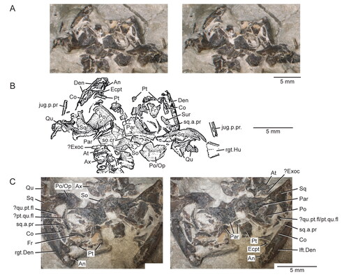
Figure 4. Virtual three-dimensional (3D) reconstruction of the skull and lower jaws of the holotype specimen (USNM PAL 722041) of Opisthiamimus gregori gen. et sp. nov. in A, dorsal, B, ventral, C, anterior, D, posterior and E, right lateral views. Colour key is at the bottom right. Solid colours indicate 3D renderings of a single bone. The mixed colours for the premaxilla and parietal indicate that for each of those bones the 3D renderings of their left and right sides were merged together to create a single, more complete version before reassembly. The left side of the skull and lower jaws is reflected from the better-preserved bones of the right side (except the vomer, pterygoid and ectopterygoid). Both left and right frontals are reassembled here.
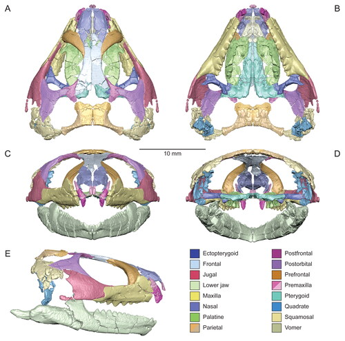
Figure 5. Virtual three-dimensional renderings of the premaxillae and maxilla of the holotype specimen (USNM PAL 722041) of Opisthiamimus gregori gen. et sp. nov. A, right premaxilla from left to right in lateral, medial, anterior and posterior views; B, portion of the right premaxilla (grey shade) preserved within an outline of a complete premaxilla based on Clevosaurus hudsoni (Fraser Citation1988, fig. 5); C, left premaxilla from left to right in lateral, medial, anterior and posterior views; D, portion of the left premaxilla (grey shade) as in B; E–H, right maxilla in E, right lateral, F, medial, G, dorsal and H, ventral views. Abbreviations: asc.pr, ascending process; cnV.for, foramen for cranial nerve five; d.pr, dorsal process; ecpt.fct, facet for the ectopterygoid; fac.pr, facial process; for, foramen; jug.fct, facet for the jugal; low.pal.fct, facet for the lower palatine process of the palatine; mx.fct, facet for the maxilla; na.fct, facet for the nasal; ntch, notch; p.pr, posterior process; pmx.fct, facet for the premaxilla; prfr.fct, facet for the prefrontal; sb, secondary bone; suborb.mar; suborbital margin; suborb.ram, suborbital ramus; t, tooth; upp.pal.fct, facet for the upper palatine process of the palatine; v.pr, ventral process.
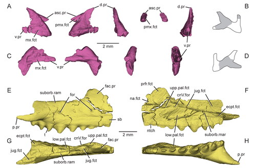
Figure 6. A referred partial skull and lower jaw (USNM PAL 720475) of Opisthiamimus gregori gen. et sp. nov. A–C, extended depth of field stereophotopairs in A, lateral, B, ventrolateral and C, medial views; D, close-up of the anterior end of the left maxilla as shown in the box in the right stereophoto in C; E–G, interpretive camera lucida drawings for A, C and B, respectively. Abbreviations: An, angular; an.fct, facet of the angular; Art, articular; Den, dentary; den.can.t, dentary successional caniniform tooth; den.co.pr, coronoid process of the dentary; den.sym.s, symphyseal surface of the dentary; ht, hatchling tooth; Jug, jugal; jug.p.pr, posterior process of the jugal; left.Pal, left palatine; lft.Mx, left maxilla; pal.t, palatine tooth; pmx.fct, facet for the premaxilla; pmx.pr, premaxillary process; Pre, prearticular; Qu, quadrate; qu.cd, quadrate condyle; r.art.pr, retroarticular process; rgt.Pal, right palatine; su, suture; Sur, surangular; wf, wear facet; ?, unidentified bone.
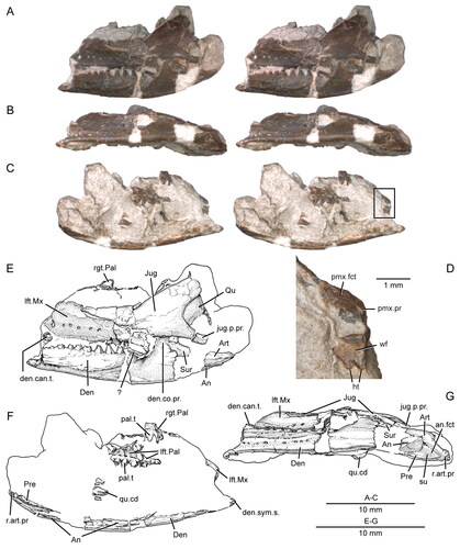
Figure 7. Virtual three-dimensional renderings of the dermal roofing bones of the skull of the holotype specimen (USNM PAL 722041) of Opisthiamimus gregori gen. et sp. nov. A, left nasal in dorsal view; B–E, right nasal in B, dorsal, C, right lateral, D, ventral and E, medial views; F–I, left frontal in F, dorsal, G, left lateral, H, ventral and I, medial views; J–M, right frontal in J, dorsal, K, right lateral, L, ventral and M, medial views; N–Q, partial left parietal in N, dorsal, O, left lateral, P, ventral and Q, medial views. Anterior is towards the top of the figure. Abbreviations: a.pr, anterior process; cr.cr, crista cranii; dep, depression; fr.fct, facet for the frontal; l.pr, lateral process; na.fct, facet for the nasal; orb.mar, margin of the orbit; par.for, parietal foramen; pofr.fct, facet for the postfrontal; prfr.fct, fact for the prefrontal; ri, ridge; sh, shelf.
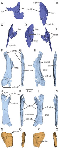
Figure 8. Virtual three-dimensional renderings of the circumorbital bones of the skull of the holotype specimen (USNM PAL 722041) of Opisthiamimus gregori gen. et sp. nov. A–D, left prefrontal in A, dorsal, B, left lateral, C, ventral and D, medial views; E–H, right prefrontal in E, dorsal, F, right lateral, G, ventral and H, medial views; I–L, right postfrontal in I, dorsal, J, right lateral, K, ventral and L, medial views; M–P, right postorbital in M, dorsal, N, right lateral, O, ventral and P, medial views; Q–T, left jugal in Q, dorsal, R, left lateral, S, ventral and T, medial views; U–X, right jugal in U, dorsal, V, right lateral, W, ventral and X, medial views. Anterior is towards the top of the figure. Abbreviations: a.pr, anterior process; bp, bony protuberance; brk, break; d.pr, dorsal process; ecpt.fct, facet for the ectopterygoid; fr.fct, facet for the frontal; gr, groove; jug.fct, facet for the jugal; l.pr, lateral process; lac.for, lacrimal foramen; ltf.mar, margin of the lower temporal fenestra; m.pr, medial process; mx.fct, facet for the maxilla; na.fct, facet for the nasal; ntch, notch; orb.mar, margin of the orbit; p.pr, posterior process; par.fct, facet for the parietal; pofr.fct, facet for the postfrontal; porb.fct, facet for the postorbital; ri, ridge; slt, slot; sq.fct, facet for the squamosal; utf.mar, margin of the upper temporal fenestra; v.pr, ventral process.
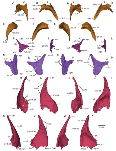
Figure 9. Details of the squamosals, articulation of the surangular and coronoid, and quadrates in the holotype specimen (USNM PAL 722041, ‘skeletal block’) of Opisthiamimus gregori gen. et sp. nov. A, extended depth of field (EDF) stereophotopair of the left posterolateral region of the skull in dorsal view; B, close-up of the surangular and coronoid bones as shown in the box in the right stereophoto in A; C, close-up of the medial half of the left squamosal as shown in the box in the left stereophoto in A; D, E, virtual three-dimensional rendering of the anteroventral portion of the left squamosal in D, left lateral and E, posteromedial views; F, EDF stereophotopair of the right posterolateral region of the skull in dorsal view; G, close-up of the possible flanges of the right quadrate and pterygoid as shown in the box in the right stereophoto in F. Abbreviations: a.pr, anterior process; brk, break; Co, coronoid; den.co.pr, coronoid process of the dentary; ecpt.v.pr, ventral process of the ectopterygoid; jug.fct, facet for the jugal; lft.jug.p.pr, posterior process of the left jugal; ltf.mar, margin of the lower temporal fenestra; m.DM.fct, facet for the mm. depressor mandibulae; ntch, notch; par.pl.pr, posterolateral process of the parietal; Po/Op, prootic/opisthotic; pt.l.fl, lateral flange of the pterygoid; pt.nk, ‘neck’ region of the pterygoid; ?pt.p.fl, possible posterior or quadrate flange of the pterygoid; Qu, quadrate; ?qu.a.fl, possible anterior or pterygoid flange of the quadrate; qu.ceph.cd, cephalic condyle of the quadrate; qu/qj.fct, facet for the quadrate/quadratojugal; rgt.Co, right coronoid; rgt.Den, right dentary; rgt.jug.p.pr, posterior process of the right jugal; ri, ridge; so.cr, supraoccipital crest; sq.a.pr, anterior process of the squamosal; sq.p.pr, posterior process of the squamosal; sq.pm.pr, posteromedial process of the squamosal; sq.v.pr, ventral process of the squamosal; sur.a.pr, anterior process of the surangular; u.dep, ‘U’-shaped depression; v.pr, ventral process; ?, unidentified bone fragment.
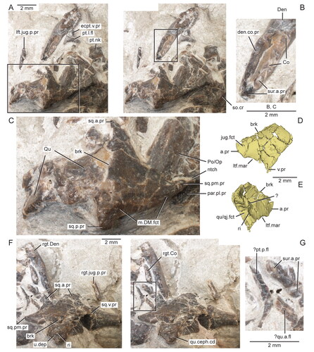
Figure 10. Virtual three-dimensional renderings of the bones of the palate of the holotype specimen (USNM PAL 722041) of Opisthiamimus gregori gen. et sp. nov. A–D, left vomer in A, dorsal, B, lateral, C, ventral and D, medial views. E, F, left pterygoid in E, dorsal and F, ventral views; G, right ectopterygoid in lateral view; H, I, left ectopterygoid in H, ventral and I, dorsal views; J–M, left palatine in J, dorsal, K, left lateral, L, ventral and M, medial views; N, displaced portion of the left palatine in anterior view; O–S, right palatine in O, dorsal, P, right lateral, Q, ventral, R, medial and S, anterior views. Arrow at J indicates where the displaced portion of the left palatine originated. Abbreviations: al.pr, anterolateral process; brk, break; ch.mar, margin of the choana; cnV.for, foramen for cranial nerve five; dep, depression; ecpt.fct, facet for the ectopterygoid; gr, groove; ipt.vac.mar, margin of the interpterygoid vacuity; jug.fct, facet for the jugal; l.fl, lateral flange; l.pr, lateral process; l.t.row, lateral tooth row of the palatine; low.mx.fct, lower maxillary facet; m.pr, medial process; mx.fct, facet for the maxilla; mx.pr, maxillary process; ntch, notch; p.pr, posterior process; pal.fct, facet for the palatine; pit, pit; pl.pr, posterolateral process; prfr.fct, facet for the prefrontal; pt.fct, facet for the pterygoid; ri, ridge; suborb.mar, margin of the suborbital fenestra; t, tooth; upp.mx.fct, upper maxillary facet; v.pr, ventral process; vo.fct, facet for the vomer.
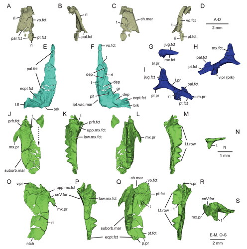
Figure 11. Virtual three-dimensional renderings of the lower jaws of the holotype (USNM PAL 722041) of Opisthiamimus gregori gen. et sp. nov. A–E, part of the right dentary in A, right lateral, B, medial, C, dorsal, D, ventral and E, anteromedial views; F, close-up of the symphyseal region of the right dentary in anteromedial view; G–K, part of the left dentary in G, anteromedial, H, left lateral, I, medial, J, dorsal and K, ventral views; L–N, right lower jaw in L, medial, M, dorsal and N, right lateral views. Arrow in L indicates the anterior extent of the angular. Abbreviations: ad.fss, adductor fossa; addt, additional tooth; An, angular; an.fct, facet for the angular; Art, articular; art.l.sulc, lateral sulcus of the articular; art.m.sulc, medial sulcus of the articular; co.pr, coronoid process; d.sym.s, dorsal symphyseal surface; Den, dentary; den.p.pr, posterior process of the dentary; ed.ri, edentulous ridge; for, foramen; ht, hatchling tooth; men.pr, mentonian process; mk.gr, Meckelian groove; ntch, notch; Pre, prearticular; psym.lam, postsymphyseal lamina; r.art.pr, retroarticular process; ri, ridge; sb, secondary bone; subd.ri, subdental ridge; succt, successional tooth; Sur, surangular; sur.a.pr, anterior process of the surangular; sur.fct, facet for the surangular; sym.s, symphyseal surface; v.sym.s, ventral symphyseal surface; wf, wear facet.
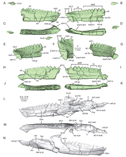
Figure 12. Extended depth of field stereophotopairs of a referred partial skull and lower jaw (USNM PAL 720475) and right dentary (USNM PAL 720476) of Opisthiamimus gregori gen. et sp. nov. A, USNM PAL 720475 in left lateral view; B–D, USNM PAL 720476 in B, lateral, C, medial and D, dorsal views. Abbreviations: an.fct, angular facet; can.t.2, second or distal successional caniniform tooth; Den, dentary; el.wf, elongate wear facet; fac.pr, facial process; for, foramen; hc.wf, half-circle-shaped wear facet; Jug, jugal; men.pr, mentonian process; mes.lab.cr, mesiolabial crest; mes.lin.cr, mesiolingual crest; mk.gr, Meckelian groove; Mx, maxilla; pmx.pr, premaxillary process; ri, ridge; sb, secondary bone; subd.ri, subdental ridge; taph.ab, taphonomic abrasion; ult.addt, ultimate or distal-most additional tooth; wax, wax; wf, wear facet; ?, unidentified bone.
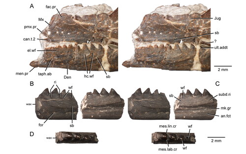
Figure 13. The maxillary teeth of Opisthiamimus gregori gen. et sp. nov. A, interpretative camera lucida drawing of the teeth of the holotype specimen (USNM PAL 722041) in occlusal view; B, C, virtual three-dimensional renderings of the right maxilla with close-ups of its teeth in the holotype specimen in B, anterolateral/mesiolabial and C, posterolateral/distolabial views. The boxes in B and C indicate the respective magnified regions to the right. Abbreviations: brk, break; can.t, successional caniniform tooth; crwn.apx, tooth crown apex; dst.car, distal carina; dst.ling.fl, distolingual flange; in.long.cr, internal longitudinal cracks of the tooth enamel; mes.car, mesial carina; succt, successional tooth; t.bs, tooth base; tri.con, triangular concavity; wf, wear facet; wr, non-occlusal wear.
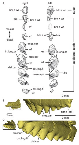
Figure 14. Dentary successional caniniform teeth of the holotype (USNM PAL 722041, ‘skull block’) of Opisthiamimus gregori gen. et sp. nov. A, extended depth of field (EDF) stereophotopair of the anterior end of the upper and lower jaws in right lateral view; B, interpretive camera lucida drawing of the anterior end of the right dentary in right lateral view; C, virtual three-dimensional rendering of the anterior end of the right dentary in anterodorsal and slightly medial views; D, EDF stereophotopair of the anterior end of the upper and lower jaws in left lateral and ventral views. Abbreviations: can.t.1, first or symphyseal successional caniniform tooth; can.t.2, second or distal successional caniniform tooth; can.t.2.imp, impression of the second or distal successional caniniform tooth; den.imp, dentary impression; dst.car, distal carina; ed.ri, edentulous ridge; for, foramen; ?ht, possible remnant of a hatchling tooth; lft.Den, left dentary; lft.den.sym.s, symphyseal surface of the left dentary; lft.Max, left maxilla; lft.Pmx, left premaxilla; men.pr, mentonian process; mes.car, mesial carina; mx.can.t, successional caniniform tooth of the maxilla; ntch, notch; psym.lam, postsymphyseal lamina; Pt, pterygoid; rgt.Den, right dentary; rgt.Max, right maxilla; rgt.Pmx, right premaxilla; sb, secondary bone; sym.s, symphyseal surface; Vo, vomer; wf, wear facet.
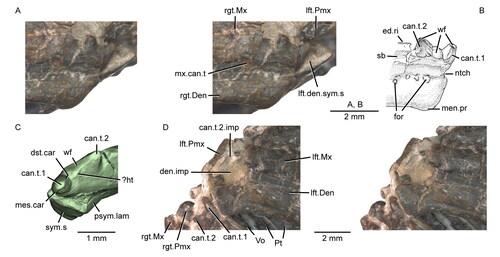
Figure 15. Dentary additional teeth of Opisthiamimus gregori gen. et sp. nov. and Opisthias rarus. A, extended depth of field (EDF) stereophotopair of the distal-most additional teeth of a referred dentary (USNM PAL 720476) of O. gregori in labial view; B, interpretive camera lucida drawing of the distal-most additional teeth of USNM PAL 720476 in occlusal view; C, EDF stereophotopair of the distal-most additional teeth of the holotype dentary (USNM V 2860) of O. rarus in labial view; D, interpretive camera lucida drawing of the distal-most additional teeth of USNM V 2860 in occlusal view; E, EDF stereophotopair of the distal-most additional teeth of the ‘paratype’ dentary (USNM V 2858) of O. rarus in labial view; F, interpretive camera lucida drawing of the distal-most additional teeth of USNM V 2858 in occlusal view; G–L, interpretive camera lucida drawings of the holotype and ‘paratype’ specimens of O. rarus, respectively, in G and H, lateral, I and J, medial, and K and L, dorsal views; M, virtual three-dimensional rendering of the holotype right dentary of O. gregori in right lateral view. C*, D*, E* and F*, regions of the tooth row shown in C, D, E and F, respectively. Arrows in B, D and F point mesially. Arrows in I, J and M indicate the anterior extent of the angular. Dashed line in I outlines the coronoid process of the dentary as illustrated in Gilmore (Citation1909, fig. 1). Abbreviations: addt, additional tooth; an.fct, facet for the angular; brk, break; co.pr, coronoid process; ed.ri, edentulous ridge; for, foramen; gr, groove or escape structure; ht, hatchling tooth; men.pr, mentonian process; mk.gr, Meckelian groove; ntch, notch; p.pr, posterior process; psym.lam, postsymphyseal lamina; ri, ridge; sb, secondary bone; succt, successional tooth; sur.fct, facet for the surangular; sym.s, symphyseal surface; t.pit, empty tooth pit; wf, wear facet.
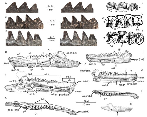
Figure 16. The palatine teeth of the holotype specimen (USNM PAL 722041, ‘skull block’) of Opisthiamimus gregori gen. et sp. nov. A, extended depth of field stereophotopair of the palatal region of the skull in left ventrolateral view; B, close-up of the isolated palatine tooth of the right palatine in occlusomedial view as shown in the box in the right stereophoto in A; C, close-up of the lateral tooth row of the right palatine in occlusolingual view as shown in the box in the left stereophoto in A; D, E, virtual three-dimensional renderings of the left palatine lateral tooth row in D, lingual, and E, occlusal views. Boxes at D and E indicate the regions of the left palatine enlarged and labeled to their left. Abbreviations: dst.lab.fl, distolabial flange; fl.surf, flattened surface; ?ht, hatchling teeth or shallowly set mature teeth; pal.can.t, caniniform tooth of the palatine; wf, wear facet.
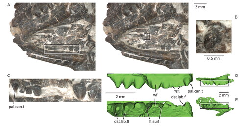
Figure 17. Partial skeleton of the holotype specimen (USNM PAL 722041, ‘skeletal block’) of Opisthiamimus gregori gen. et sp. nov. A, digital photograph in dorsal view; B, interpretive camera lucida drawing for A. Abbreviations: At, atlas or presacral vertebra no. 1; auto, autopodium; Ax, axis or presacral vertebra no. 2; bra, braincase; Cv5, cervical or presacral vertebra no. 5; cv.rb4, cervical rib no. 4; cv.rb7, cervical rib no. 7; Den, dentary; Fe, femur; Hu, humerus; I, ilium; Jug, jugal; ?Os, possible osteoderm; Par, parietal; Ph, phalanx; Psv15, presacral vertebra no. 15; Psv-7, seventh from last presacral vertebra; psv.rb14, presacral rib no. 14; psv.rb-6, sixth from last presacral rib; Psv.ult, ultimate or last presacral vertebra; Qu, quadrate; Ra, radius; Rb, rib; Sav, sacral vertebra; ?Sc, possible scapula; Sq, squamosal; Ul, ulna.
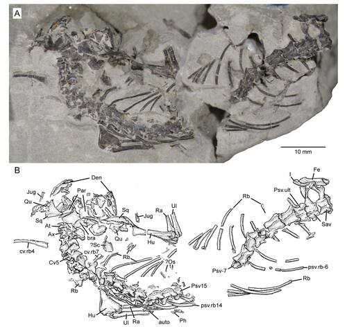
Figure 18. Anterior cervical vertebrae of Opisthiamimus gregori gen. et sp. nov. A, extended depth of field (EDF) stereophotopair of the anterior cervical series in left lateral view of the holotype specimen (USNM PAL 722041, ‘skeletal block’); B, interpretive camera lucida drawing for A; C–G, EDF stereophotopairs of a referred partial atlas/axis complex (USNM PAL 720479) in C, dorsal, D, left lateral, E, ventral, F, anterior and G, posterior views; H–L, interpretive camera lucida drawings for C–G, respectively. Abbreviations: accp, accessory process; At, atlas vertebra; at.a.pr, anterior process of the atlas; At.cen (od), atlas centrum or odontoid process; Ax, axis vertebra; ax.cen, centrum of the axis vertebra; ax.n.a, neural arch of the axis vertebra; bp, bony protuberance; brk, break; cp.tb, capitular protuberance of the tuberculum; Cv5, fifth cervical vertebra; cv.rb5, cervical rib no. 5; dep, depression; ?Exoc, possible exoccipital; Ic2, second intercentrum; int.zyg.ri, interzygapophyseal ridge; n.a., neural arch; nsp, neural spine; p.co, posterior cotyle; pozyg, postzygapophysis; prezyg, prezygapophysis; su, suture; syn, synapophysis; tb, tuberculum.
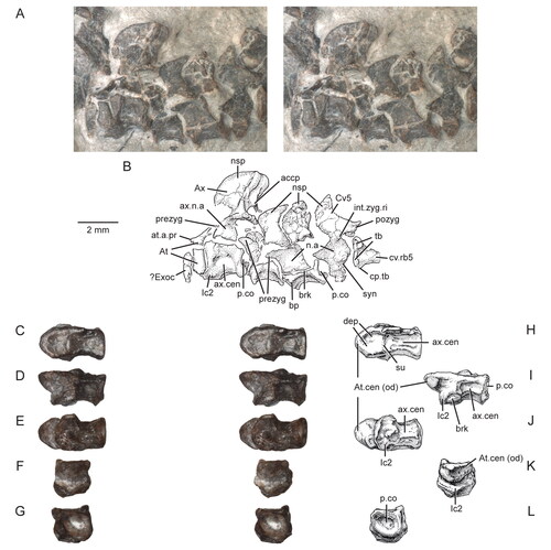
Figure 19. Presacral and sacral vertebrae of the holotype specimen (USNM PAL 722041, ‘skeletal block’) of Opisthiamimus gregori gen. et sp. nov. A, extended depth of field (EDF) stereophotopair of presacral vertebrae 11 to 15 in dorsal view; B, EDF stereophotopair of the seventh to third from last presacral vertebrae in dorsal view; C, EDF stereophotopair of the posterior-most four presacral vertebrae and the sacral vertebrae in dorsal view; D, camera lucida drawing of the skeleton in dorsal view with boxes labeled A*, B*, and C* indicating the regions shown in A–C, respectively. Abbreviations: ?G, possible gastralium; int.zyg.ri, interzygapophyseal ridge; lft.Ra, left radius; lft.Ul, left ulna; ?Os, possible osteoderm; Ph, phalanx; Psv11, presacral vertebra no. 11; Psv15, presacral vertebra no. 15; Psv-3, third from last presacral vertebra; Psv-6, sixth from last presacral vertebra; Psv.ult, ultimate or last presacral vertebra; Rb, rib; ri, ridge; Sav1, sacral vertebra no. 1; syn, synapophysis; zys, zygosphene.
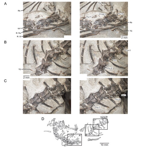
Figure 20. Sacral vertebrae of the holotype specimen (USNM PAL 722041, ‘skeletal block’) of Opisthiamimus gregori gen. et sp. nov. A, extended depth of field (EDF) stereophotopair of the posterior-most presacral and sacral vertebrae in dorsal view; B, interpretive camera lucida drawing for A; C, interpretive camera lucida drawing for D; D, EDF stereophotopair of the posterior-most presacral and sacral vertebrae in posterolateral and dorsal views. Abbreviations: brk, break; dia, diapophysis; Fe, femur; I, ilium; in.tr, internal trochanter; lam, lamina; n.cnl, neural canal; nsp, neural spine; p.co, posterior cotyle; p.pr, posterior process; para, parapophysis; pozyg, postzygapophysis; prezyg, prezygapophysis; Psv-3, third from last presacral vertebra; Psv.ult, ultimate or last presacral vertebra; px.hd, proximal head; Rb, rib; sa.rb, sacral rib; Sav1, sacral vertebra no. 1; Sav2, sacral vertebra no. 2; ?, unidentified bone.
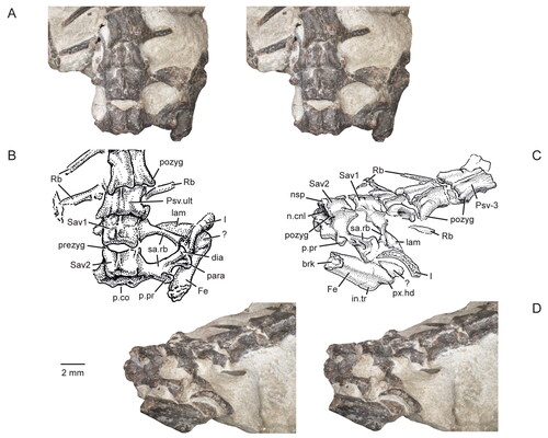
Figure 21. Caudal vertebra of the holotype specimen (USNM PAL 722041) of Opisthiamimus gregori gen. et sp. nov. A, extended depth of field (EDF) stereophotopair in left lateral view; B, interpretive camera lucida drawing of A; C, EDF stereophotopair in right lateral view; D, interpretive camera lucida drawing of C; E, EDF stereophotopair in dorsal view; F, interpretive camera lucida drawing of E; G–I, interpretive camera lucida drawings in G, ventral, H, anterior and I, posterior views. Abbreviations: a.co, anterior cotyle; brk, break; cen, centrum; n.cnl, neural canal; nsp, neural spine; p.co, posterior cotyle; pozyg, postzygapophysis; prezyg, prezygapophysis; subc.ri, subcentral ridge; tr.pr, transverse process.
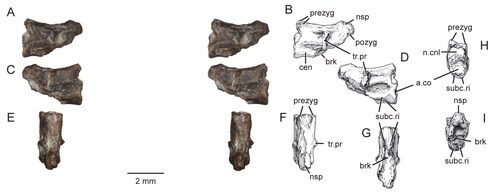
Figure 22. Forelimbs and a portion of the axial skeleton of the holotype specimen (USNM PAL 722041, ‘skeletal block’) of Opisthiamimus gregori gen. et sp. nov. A, extended depth of field (EDF) stereophotopair of the exposed left forelimb and several presacral vertebrae and associated ribs in dorsal view; B, interpretive camera lucida drawing for A; C, interpretive camera lucida drawing for D; D, EDF stereophotopair of the exposed right forelimb and adjacent bones of the skull in dorsal view. Anterior is to the left in A and B and towards the top in C and D. Abbreviations: auto, autopodium; ect, ectepicondyle; ect.for, ectepicondylar foramen; ent, entepicondyle; ent.for, entepicondylar foramen; ?G, possible gastralia; Hu, humerus; ol, olecranon process; Psv10, presacral vertebra no. 10; Psv13, presacral vertebra no. 13; Qu, quadrate; Ra, radius; Rb, rib; rgt.jug.p.pr, posterior process of the right jugal; Sq, squamosal; tr, trochlea; tu, tuberculum; Ul, Ulna; v.dep, ‘V’-shaped depression.
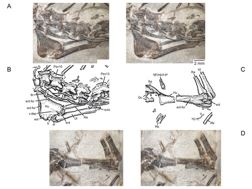
Figure 23. Distal radius and ulna, carpus and manus of the holotype specimen (USNM PAL 722041, ‘hand block’) of Opisthiamimus gregori gen. et sp. nov. (A–C) and Sphenodon punctatus (D, E). A, extended depth of field stereophotopair of the right distal forearm, carpus and manus in ventral (palmar) view; B, interpretive camera lucida drawing for A; C, virtual three-dimensional rendering of the right distal forearm, carpus and manus in dorsal view; D, left distal forearm, carpus and manus (reflected for ease of comparison) in dorsal view; E, carpus based on S. punctatus in D showing which elements are likely preserved in O. gregori (dark grey). The solid lines represent the carpals that most likely are present; the thick dashed line represents the presence only tentatively. The illustration in D is from an original, unpublished inkwash and pencil drawing made for O. C. Marsh in March, 1886 (artist unknown), and housed in the Department of Paleobiology collections at the NMNH. Abbreviations: C, carpus; col.lig.fos, collateral ligament fossa; d1–d5, distal carpals one to five; gr, groove; I–V, digits one to five; ?Int, possible intermedium; lC, lateral centrale; mC, medial centrale; Mc, metacarpal; ?Os, possible osteoderm; P, pisiform; Ph, phalanx; Ra, radius; Re, radiale; Ue, ulnare; Ul, ulna; Un, ungual; un.flx.tu, flexor tubercle of the ungual.
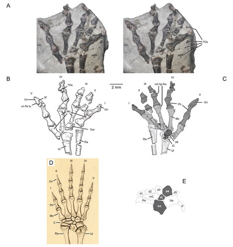
Figure 24. Consensus trees based on maximum parsimony (A–C) and Bayesian (D) analyses with Opisthiamimus gregori gen. et sp. nov. and Leptorhynchia taxon nov. shown in bold font. A, strict consensus of 24 most parsimonious trees (MPTs; 436 steps each, consistency index [CI] = 0.3991, retention index [RI] = 0.6421) with clade names at their respective nodes; B, Adams consensus of 24 MPTs (426 steps each, CI = 0.4085, RI = 0.6557) with Bremer (>1) and bootstrap (>50) support values provided above and below each node, respectively, that is supported as such (all nodes with these values also are recovered in the strict consensus in A; C, strict consensus of 24 MPTs following the iterPCR protocol demonstrating the alternative positions of the most phylogenetically unstable taxa; D, 50% majority rule consensus tree with posterior probabilities provided.
![Figure 24. Consensus trees based on maximum parsimony (A–C) and Bayesian (D) analyses with Opisthiamimus gregori gen. et sp. nov. and Leptorhynchia taxon nov. shown in bold font. A, strict consensus of 24 most parsimonious trees (MPTs; 436 steps each, consistency index [CI] = 0.3991, retention index [RI] = 0.6421) with clade names at their respective nodes; B, Adams consensus of 24 MPTs (426 steps each, CI = 0.4085, RI = 0.6557) with Bremer (>1) and bootstrap (>50) support values provided above and below each node, respectively, that is supported as such (all nodes with these values also are recovered in the strict consensus in A; C, strict consensus of 24 MPTs following the iterPCR protocol demonstrating the alternative positions of the most phylogenetically unstable taxa; D, 50% majority rule consensus tree with posterior probabilities provided.](/cms/asset/fd704d2e-8180-471c-a185-562a5825dea3/tjsp_a_2093139_f0024_b.jpg)
Figure 25. A time-calibrated phylogeny of Rhynchocephalia (and outgroups) based on the strict consensus presented in . The thick bars and boxes at the branch tips represent the temporal ranges for each species. The Morrison Formation taxa are emphasized by the enlarged bold font and black-bordered white boxes. Boundary dates are based on Walker et al. (Citation2018). The three-dimensional reconstructed skull of Opisthiamimus gregori gen. et sp. nov. is shown in oblique anterodorsolateral view. Abbreviations: Ac, Acrosphenodontia; Cl, Clevosauridae; E, Early; Ei, Eilenodontinae; Eoc, Eocene; Eu, Eusphenodontia; Gua, Guadalupian; L, Late; Lep, Lepidosauria; LEPT, Leptorhynchia taxon nov.; Lo, Lopingian; M, Middle; Ma, million years ago: Mio, Miocene; N, Neogene; Neo, Neosphenodontia; O, Oligocene; Pal, Paleocene; Pl, Pleurosauridae; R, Rhynchocephalia; S, Sphenodontidae; Sa, Saphaeosauridae; Sp, Sphenodontia; *, Pliocene, Pleistocene and Holocene.
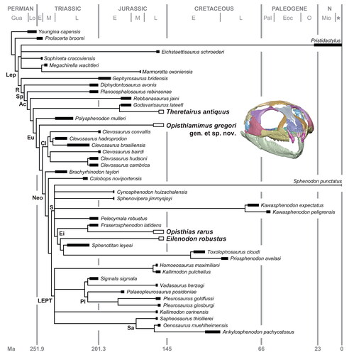
Figure 26. Distributions of some of the major synapomorphies in Rhynchocephalia as mapped onto the maximum parsimony strict consensus tree, including many that are hypothesized to be associated with proal and orthal jaw movements (e.g. characters 46, 53, 59, 60, 63, 90, 91, 93). Major clade nodes are indicated with coloured circles. Bold numbers above nodes represent those listed in the full apomorphy list for the strict consensus (see Supplemental material). The coloured circles and boxes at and below the nodes correspond to those bracketing the major clades at the top of the figure. The horizontal rectangles on the stem below each major node indicate some or all of the unambiguous or ambiguous character states supporting that node; ambiguous character states are denoted with an asterisk (*). Black rectangles = cranial, lower jaw or dental characters. White-filled rectangles = axial skeleton characters. Grey-filled rectangles = appendicular skeleton characters. Numbers and numbers in parentheses in the form of X(Y) and next to the rectangles represent the character and character state numbers presented in the character and apomorphy lists (see Supplemental material). Abbreviations: Cy., Cynosphenodon; H., Homoeosaurus; Pe., Pelecymala; Si., Sigmala; Sp., Sphenovipera; V., Vadasaurus.
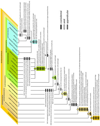
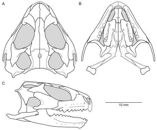
Figure 27. Line drawing reconstruction of the skull (minus the braincase) and lower jaws of Opisthiamimus gregori gen. et sp. nov. based on observations of the holotype and referred specimens (USNM PAL 720475, 720476, 722041) and our three-dimensional reconstructions (e.g. ; Supplemental material, Fig. S4). A, dorsal view; B, ventral view; C, right lateral view. Light grey represents cranial openings (i.e. orbit, choana, upper and lower temporal, infraorbital and suborbital fenestrae, external nares) and foramina (i.e. parietal, mandibular); dashed lines represent hypothesized contours.