Figures & data
Figure 1. The geological setting of NJSM GP12145. A, outline of the USA with the state of New Jersey highlighted in black overlaying map with New Jersey highlighted in grey. Coral highlighting the combined Hornerstown and Vincentown formations. Fossil locality, the Jean and Ric Edelman Fossil Park, in Sewell marked with a black star. Adapted from Owens et al. (Citation1999) and Dalton et al. (Citation2014). B, stratigraphical section at the Jean and Ric Edelman Fossil Park. NJSM GP12145 derives from the Danian portion of the Main Fossiliferous Layer of the Hornerstown Formation. The combined Hornerstown and Vincentown formations are highlighted in coral. Adapted from Staron et al. (Citation2001).
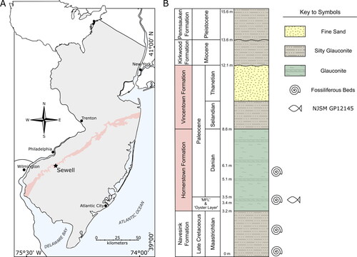
Figure 2. Photogrammetric model of the skull of †Iridopristis parrisi, gen. et sp. nov., holotype (NJSM GP12145), Hornerstown Formation, early Paleocene (Danian), New Jersey, USA.
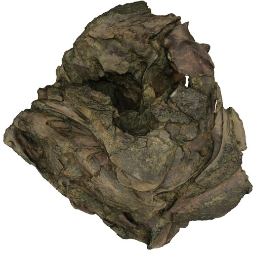
Figure 3. Skull and abdomen of †Iridopristis parrisi in left lateral view. Holotype (NJSM GP12145), Hornerstown Formation, early Paleocene (Danian), New Jersey, USA. A, specimen photograph and B, rendered µCT model. Skeletal regions highlighted as follows: neurocranium (pink), suspensorium (purple), circumorbitals (coral), jaws (light blue), opercles (light orange), ventral hyoid (light green), gill skeleton (dark green), pectoral girdle (yellow), abdominal scales (dark orange), vertebral column (red). Arrow indicates anatomical anterior. Scale bar represents 5 cm.
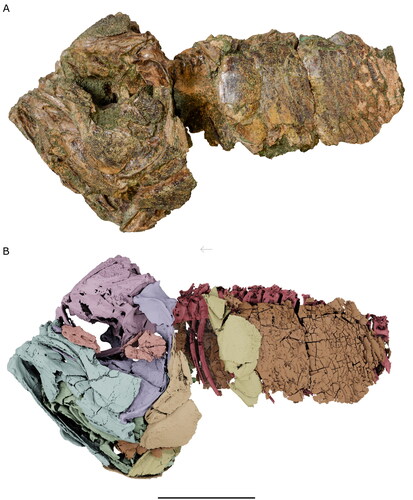
Figure 4. Skull and abdomen of †Iridopristis parrisi in right lateral view. Holotype (NJSM GP12145), Hornerstown Formation, early Paleocene (Danian), New Jersey, USA. A, specimen photograph and B, rendered µCT model. Skeletal regions highlighted as follows: neurocranium (pink), suspensorium (purple), circumorbitals (coral), jaws (light blue), opercles (light orange), ventral hyoid (light green), gill skeleton (dark green), pectoral girdle (yellow), scales (dark orange), vertebral column (red). Arrow indicates anatomical anterior. Scale bar represents 5 cm.
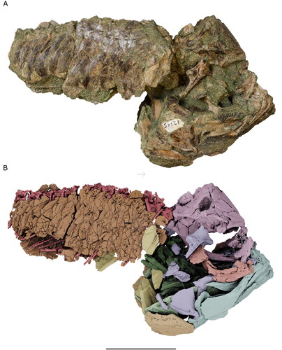
Figure 5. Referred specimens of †Iridopristis parrisi. NJSM GP12381, Hornerstown Formation, early Paleocene (Danian), New Jersey, USA, comprising remains of two individuals preserving portions of neurocrania, pectoral girdle, opercular series and vertebral column. Upper row depicts the left side of specimens, lower row depicts the right side of specimens. A, individual preserving neurocranium including intact supraoccipital crest, fragments of left hyomandibula, right operculum, both posttemporals, and the first three vertebral centra; and B, specimen preserving neurocranium including fragmentary supraoccipital crest, right operculum, both posttemporals, and the first three vertebral centra. Abbreviations: c, centra; fr, frontal; n, neurocranium; op, opercular; pas, parasphenoid; pt, posttemporal; soc, supraoccipital. Arrows indicate anatomical anterior. Scale bar represents 5 cm.
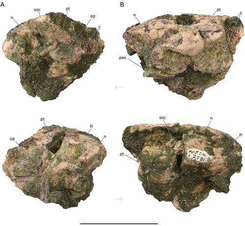
Figure 6. Neurocranium of †Iridopristis parrisi. Holotype (NJSM GP12145), Hornerstown Formation, early Paleocene (Danian), New Jersey, USA. Rendered µCT model in A, right lateral, B, posterior, C, dorsal and D, ventral views. Arrows indicate anatomical anterior. Scale bar represents 5 cm.
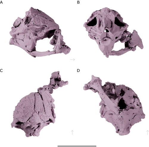
Figure 7. Neurocranium of †Iridopristis parrisi. Holotype (NJSM GP12145), Hornerstown Formation, early Paleocene (Danian), New Jersey, USA. Line drawings of rendered µCT models in A, right lateral, B, posterior, C, dorsal and D, ventral views. Abbreviations: boc, basioccipital; epo, epioccipital; exo, exoccipital; fr, frontal; hyfa, anterior facet for hyomandibula; hyfd, dorsal facet for hyomandibula; let, lateral ethmoid; met, mesethmoid; pa, parietal; pas, parasphenoid; pro, prootic; pto, pterotic; pts, pterosphenoid; soc, supraoccipital; v, vomer. Arrows indicate anatomical anterior. Scale bar represents 5 cm.
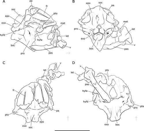
Figure 8. Right upper and lower jaws of †Iridopristis parrisi. Holotype (NJSM GP12145), Hornerstown Formation, early Paleocene (Danian), New Jersey, USA. Rendered µCT models of A, upper jaw in lateral view (maxilla in light blue, premaxilla in dark blue), B, upper jaw in mesial view, C, lower jaw in lateral view, and D, lower jaw in mesial view. Abbreviations: a, anguloarticular; d, dentary; m, maxilla; mpf, posterior facet of maxilla; pm, premaxilla; ra, retroarticular; smf, facet for supramaxillae. Arrows indicate anatomical anterior. Scale bar represents 5 cm.
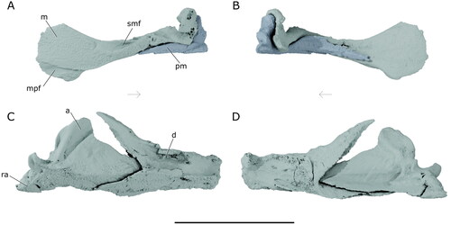
Figure 9. Suspensorium of †Iridopristis parrisi. Holotype (NJSM GP12145), Hornerstown Formation, early Paleocene (Danian), New Jersey, USA. Rendered µCT model in A, lateral and B, mesial views. Abbreviations: ecp, ectopterygoid; enp, endopterygoid; h, hyomandibula; mpt, metapterygoid; pal, palatine; q, quadrate; sym, symplectic. Arrows indicate anatomical anterior. Scale bar represents 5 cm.
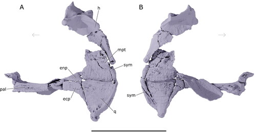
Figure 10. Left preopercle and interopercle of †Iridopristis parrisi. Holotype (NJSM GP12145), Hornerstown Formation, early Paleocene (Danian), New Jersey, USA. Rendered µCT model in A, lateral and B, mesial views. Photographs of operculum in C, lateral and D, mesial views. Abbreviations: iop, interopercle; pop, preopercle. Arrows indicate anatomical anterior. Scale bar represents 5 cm for panels A and B, and 1 cm for panels C and D.
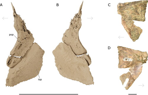
Figure 11. Left ventral hyoid apparatus of †Iridopristis parrisi. Holotype (NJSM GP12145), Hornerstown Formation, early Paleocene (Danian), New Jersey, USA. Rendered µCT models showing the A, lateral and B, mesial views of the hypohyals, ceratohyals, interhyal, and branchiostegals, and the C, urohyal in lateral and D, ventral view. Abbreviations: br, branchiostegals; cha, anterior ceratohyal; chp, posterior ceratohyal; hhd, dorsal hypohyal; hhv, ventral hypohyal; ih, interhyal; uh, urohyal. Arrows indicate anatomical anterior. Scale bar represents 5 cm.
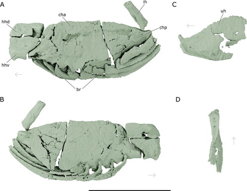
Figure 12. Gill skeleton of †Iridopristis parrisi. Holotype (NJSM GP12145), Hornerstown Formation, early Paleocene (Danian), New Jersey, USA. Rendered µCT models of A, complete gill skeleton in lateral view, including the dorsal and ventral gill skeleton and associated gill rakers, B, ventral gill skeleton in dorsal view, C, dorsal gill skeleton in ventral view. Ventral gill skeleton in green, dorsal gill skeleton in olive, gill rakers in lime. Abbreviations: bb, basibranchials; cb, ceratobranchials; eb, epibranchials; hb, hypobranchials; pb, pharyngobranchials; rk, gill rakers. Arrows indicate anatomical anterior. Scale bar represents 5 cm.
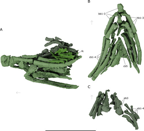
Figure 13. Pectoral girdle of †Iridopristis parrisi. Holotype (NJSM GP12145), Hornerstown Formation, early Paleocene (Danian), New Jersey, USA. Rendered µCT model in A, left lateral and B, right lateral views. Abbreviations: cl, cleithrum; co, coracoid; pcl, postcleithrum; scl, supracleithrum. Arrows indicate anatomical anterior. Scale bar represents 5 cm.
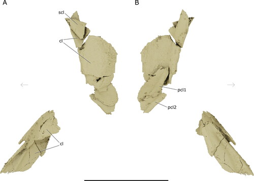
Figure 14. Squamation of †Iridopristis parrisi. Holotype (NJSM GP12145), Hornerstown Formation, early Paleocene (Danian), New Jersey, USA. Photographs of the A, cheek and B, abdominal squamation. Arrows indicate anatomical anterior. Scale bars represent 1 cm.
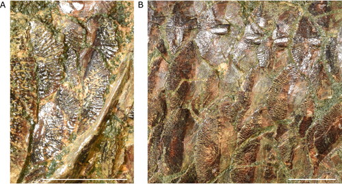
Figure 15. Axial skeleton of †Iridopristis parrisi. Holotype (NJSM GP12145), Hornerstown Formation, early Paleocene (Danian), New Jersey, USA. Shown in left lateral view. Abbreviations: c, centra; en, epineural; hs, haemal spines; r, ribs. Arrow indicates anatomical anterior. Scale bar represents 5 cm.
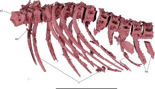
Figure 16. Variation in saccular otolith morphology among holocentroids and outgroups. Rendered µCT models of saccular otoliths as a visual explanation of states in Character 81 for A, †Iridopristis parrisi (NJSM 12145), B, Centroberyx affinis (UMMZ 216732), C, Myripristis murdjan (UMMZ 185696), and D, Sargocentron rubrum (UMMZ 245614). Arrow indicates anatomical anterior. Images not to scale.
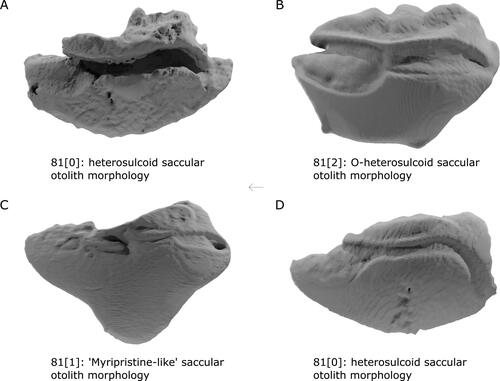
Figure 17. Variation in anguloarticular morphology in holocentroids and outgroups. Rendered µCT models of lower jaws as a visual explanation of character states in Character 82 for A, †Iridopristis parrisi (NJSM 12145), B, Centroberyx affinis (UMMZ 216732), C, Myripristis murdjan (UMMZ 185696), and D, Sargocentron rubrum (UMMZ 245614). White arrows in (C) and (D) indicate laminar flange anterior to jaw joint. Central arrow indicates anatomical anterior. Images not to scale.
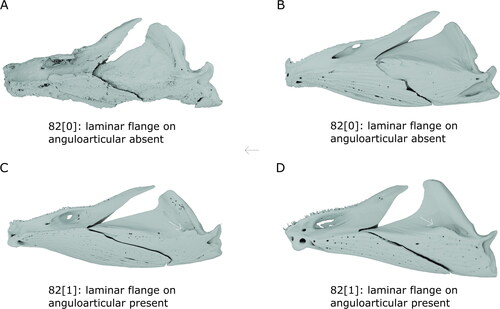
Figure 18. Holocentroid relationships inferred from a maximum parsimony analysis of morphological data. A, strict consensus of 588 most parsimonious trees with tree length = 112 steps. Circles at nodes indicate bootstrap support values between 50 (white) and 100 (black). Bremer support indices indicated by numbers adjacent to nodes. †Iridopristis parrisi in bold. B, agreement subtree of 588 most-parsimonious trees with length = 112 steps. Letters at nodes indicate character states supporting clades of interest. Character states for characters 1–80 described in detail in Dornburg et al. (Citation2015). Characters 81 and 82 new to this paper. Character-state optimizations keyed to characters at nodes: (a) 10[1], last pleural rib expanded in mediolateral plane; 11[1], second ural centrum fused to the combined first ural centrum and first preural centrum; 12[1], loss of hypural six. (b) 2[1], transverse crest of the supraoccipital; 3[1], separate opening of the orbital branch of the supraorbital sensory canal; 6[1], ventrolateral wings of the parasphenoid; 8[1], anterior ceratohyal with no foramen (closed ontogenetically); 9[1], deep notches on the ventral margin of the anterior ceratohyal to accommodate branchiostegals; 27[1], alveolar platform expanded near the symphysis to overhang the lateral margin of the dentary; 28[1], concave premaxillary tooth gap at the mesial margin. (c) 82[1], dorsally projecting lamina anterior to the jaw joint on the anguloarticular. (d) 50[1], lateral connection of the otic bulla to the swim bladder; 51[1], prootic with an elongate process projecting laterally ventral to the pars jugularis foramen. (e) 30[0], loss of teeth on the ectopterygoid; 31[3], five spinous procurrent caudal rays in upper lobe and four in lower lobe; 35[0] 16 preural caudal vertebrae. (f) 19[1], ascending process of the premaxilla as long as or longer than the alveolar process; 20[1], postmaxillary process of the premaxilla anteroposteriorly short and dorsoventrally high; 21[1], depth to length ratio of first two circumorbitals between 1:4.5 and 1:7; 22[1], shaft of the maxilla dorsoventrally deep and anteroposteriorly short; 27[0], loss of alveolar platform expansion near the symphysis to overhang the lateral margin of the dentary; 28[0], loss of convex premaxillary tooth gap at the mesial margin; 76[1], first dorsal fin pterygiophore inserts into a pocket on the anterior margin of the neural spine of the second vertebra; 78[1], lachrymal with a wide-based, ventrally directed, and short spine; 79[1], single preopercular spine in adults.
![Figure 18. Holocentroid relationships inferred from a maximum parsimony analysis of morphological data. A, strict consensus of 588 most parsimonious trees with tree length = 112 steps. Circles at nodes indicate bootstrap support values between 50 (white) and 100 (black). Bremer support indices indicated by numbers adjacent to nodes. †Iridopristis parrisi in bold. B, agreement subtree of 588 most-parsimonious trees with length = 112 steps. Letters at nodes indicate character states supporting clades of interest. Character states for characters 1–80 described in detail in Dornburg et al. (Citation2015). Characters 81 and 82 new to this paper. Character-state optimizations keyed to characters at nodes: (a) 10[1], last pleural rib expanded in mediolateral plane; 11[1], second ural centrum fused to the combined first ural centrum and first preural centrum; 12[1], loss of hypural six. (b) 2[1], transverse crest of the supraoccipital; 3[1], separate opening of the orbital branch of the supraorbital sensory canal; 6[1], ventrolateral wings of the parasphenoid; 8[1], anterior ceratohyal with no foramen (closed ontogenetically); 9[1], deep notches on the ventral margin of the anterior ceratohyal to accommodate branchiostegals; 27[1], alveolar platform expanded near the symphysis to overhang the lateral margin of the dentary; 28[1], concave premaxillary tooth gap at the mesial margin. (c) 82[1], dorsally projecting lamina anterior to the jaw joint on the anguloarticular. (d) 50[1], lateral connection of the otic bulla to the swim bladder; 51[1], prootic with an elongate process projecting laterally ventral to the pars jugularis foramen. (e) 30[0], loss of teeth on the ectopterygoid; 31[3], five spinous procurrent caudal rays in upper lobe and four in lower lobe; 35[0] 16 preural caudal vertebrae. (f) 19[1], ascending process of the premaxilla as long as or longer than the alveolar process; 20[1], postmaxillary process of the premaxilla anteroposteriorly short and dorsoventrally high; 21[1], depth to length ratio of first two circumorbitals between 1:4.5 and 1:7; 22[1], shaft of the maxilla dorsoventrally deep and anteroposteriorly short; 27[0], loss of alveolar platform expansion near the symphysis to overhang the lateral margin of the dentary; 28[0], loss of convex premaxillary tooth gap at the mesial margin; 76[1], first dorsal fin pterygiophore inserts into a pocket on the anterior margin of the neural spine of the second vertebra; 78[1], lachrymal with a wide-based, ventrally directed, and short spine; 79[1], single preopercular spine in adults.](/cms/asset/2f7030f3-fbd2-4b24-bf4d-7ffa21eb3f16/tjsp_a_2168571_f0018_b.jpg)
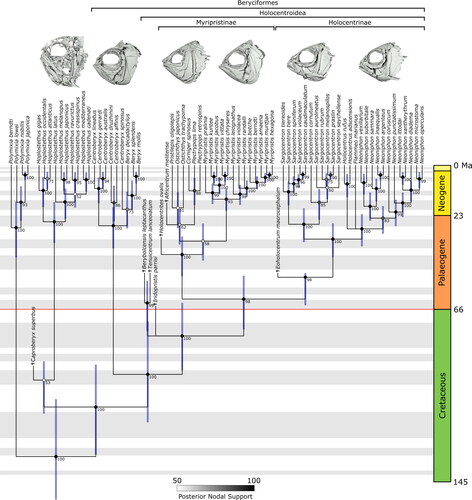
Figure 19. Maximum clade credibility tree for a total-evidence analysis of holocentroid interrelationships. Bars at nodes represent 95% HPD (highest posterior density) for node age. Nodal supported indicated as posterior probabilities between 50 (white) and 100 (black). The Cretaceous–Palaeogene boundary is highlighted in red.