Figures & data
Figure 1. Radiodont phylogeny. A, strict consensus of nine shortest cladograms under equal character weights; numbers at nodes are jackknife frequencies >50%. B, single best fit cladogram under implied weights (k = 3); numbers at nodes are G/C values >50%. Colours indicate clades: Euarthropoda (yellow), Hurdiidae (purple), Amplectobeluidae (blue), Anomalocarididae (green), and Tamisiocarididae sensu stricto (pink).
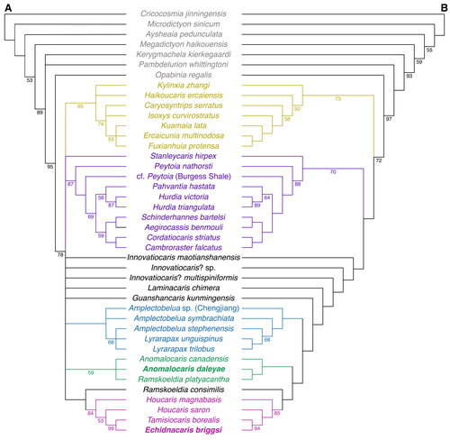
Figure 2. Anomalocaris daleyae sp. nov. Holotype SAMA P51398a. Paired frontal appendages and oral cone. A, photograph. B, camera lucida drawing (grey lines correspond to cuticle wrinkles). Abbreviations: BEn, base endite; Cp1–Cp9, claw podomeres 1–9; ds, dorsal spine; En1–9, endites of claw podomeres 1–9; ts, terminal spine on Cp13. Scale bars: 10 mm.
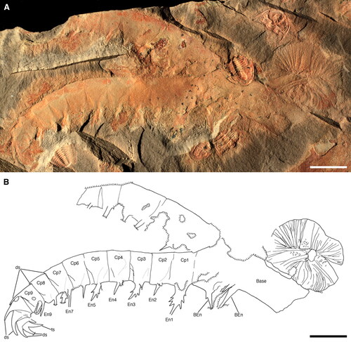
Figure 3. Anomalocaris daleyae sp. nov. Holotype SAMA P51398a. Details of frontal appendage and overview of oral cone. A, endite 1 (En1). B, endite 3 (En3). C, endite 5 (En5). D, distal part of frontal appendage. E, oral cone. Arrowheads in A–C indicate auxiliary spines, anterior to left, posterior to right. Abbreviations: Cp9–Cp12, claw podomeres 9–12; ds, dorsal spine; ts, terminal spine on Cp13. Scale bars: A–D = 2 mm; E = 5 mm.
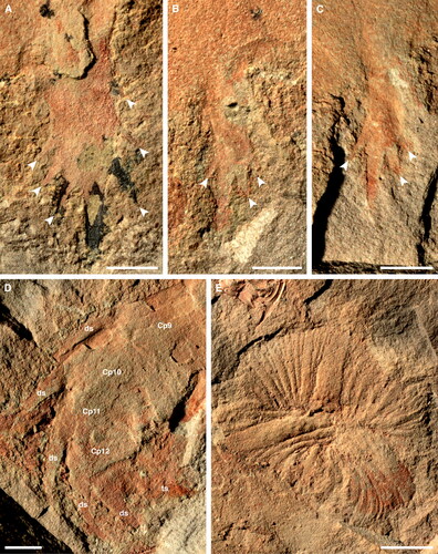
Figure 4. Anomalocaris daleyae sp. nov. Paratype SAMA P54844a, b. Paired frontal appendages and partial oral cone. A, B, P54844a. Photograph and camera lucida drawing, respectively. C, D, P54844b. Photograph and camera lucida drawing, respectively. Abbreviations: BEn, base endite; Cp1–Cp11, claw podomeres 1–11; ds, dorsal spine; En1–7, endites of claw podomeres 1–7; oc, oral cone. Scale bars: 10 mm.
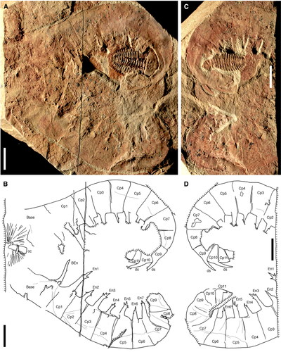
Figure 5. Anomalocaris daleyae sp. nov. Paratype SAMA P55619b. Frontal appendage. A, B, photograph and camera lucida drawing, respectively. C, endite 5 (En5). D, endite 7 (En7). Arrowheads in C, D indicate auxiliary spines; anterior to left, posterior to right. Abbreviations: Cp1–Cp11, claw podomeres 1–11; ds, dorsal spine; En1–9, endites of claw podomeres 1–9. Scale bars: A, B = 10 mm; C, D = 3 mm.
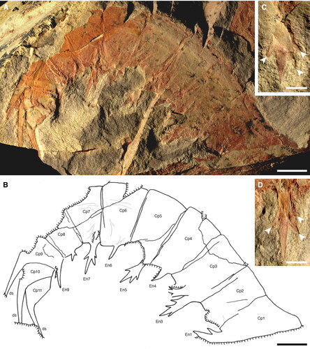
Figure 6. Anomalocaris daleyae sp. nov. A, B, paratype SAMA P15374b. Frontal appendage. A, overview. B, detail of proximal and distal parts of enrolled appendage; arrowheads indicate auxiliary spines. C, SAMA P54915. Distal part of frontal appendage, showing four dorsal spines. D, E, SAMA P54874a, b, respectively. Oral cone. Abbreviations: BEn, base endite; Cp1–Cp10, claw podomeres 1–10; ds, dorsal spine; En1, endite of claw podomere 1. Scale bars: A = 10 mm; B–E = 5 mm.
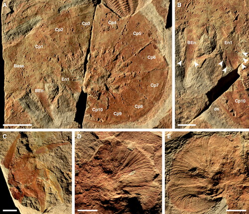
Figure 7. Reconstructions of Emu Bay Shale radiodont frontal appendages. A, B, Anomalocaris daleyae sp. nov. A, entire appendage. B, oblique cross-section of first claw podomere (Cp1) showing enlarged paired endites, each bearing three pairs of auxiliary spines. C, D, Echidnacaris briggsi (Nedin, Citation1995). C, entire appendage. D, oblique cross-section of typical claw podomere in middle portion of appendage showing paired endites bearing both auxiliary spines and spinules.
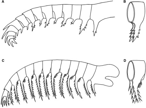
Figure 8. Echidnacaris briggsi (Nedin, Citation1995) comb. nov. Holotype SAMA P40180a, b. Frontal appendage. A, overview of part. B, detail of distal end of appendage, including podomeres 12 and 13, and En11. C, camera lucida drawing (incorporating information from counterpart). D, overview of counterpart (flipped horizontally to facilitate comparison). Abbreviations: BEn, base endite; Cp2—Cp12, claw podomeres 2–12; ds, dorsal spine; En1–11, endites of claw podomeres 1–11; sp, spinules; ts, terminal spines on Cp13. Scale bars: A, C, D = 20 mm; B = 5 mm.
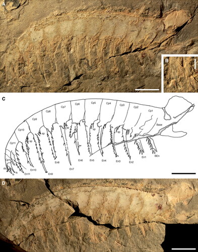
Figure 9. Echidnacaris briggsi (Nedin, Citation1995) comb. nov. SAMA P54876. Frontal appendage. A, B, P54876a, overview. Photograph and camera lucida drawing, respectively. C, P54876b, detail of counterpart, showing well-preserved endites (En2–8) with auxiliary spines in epirelief. Abbreviations: BEn, base endite; Cp1—Cp11, claw podomeres 1–11; En1–11, endites of claw podomeres 1–11; sp, spinules. Scale bars: A, B = 20 mm; C = 10 mm.
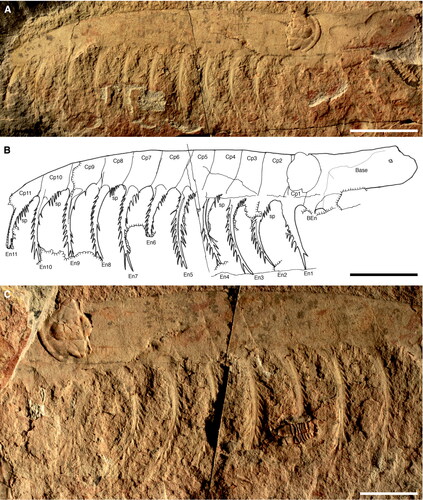
Figure 10. Echidnacaris briggsi (Nedin, Citation1995) comb. nov. SAMA P48975a. Frontal appendage. A, B, photograph and camera lucida drawing, respectively. C, detail of BEn and En1. Abbreviations: BEn, base endite; Cp1–Cp9, claw podomeres 1–9; En1–5, endites of claw podomeres 1–5; sp, spinules. Scale bars: A, B = 20 mm; C = 10 mm.
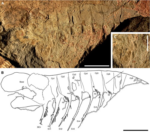
Figure 11. Echidnacaris briggsi (Nedin, Citation1995) comb. nov. SAMA P54790a. Frontal appendage. A, B, photograph and camera lucida drawing, respectively. Abbreviations: Cp5—Cp13, claw podomeres 5–13; ds, dorsal spine; En5–13, endites of claw podomeres 5–13; sp, spinules. Scale bars: 10 mm.
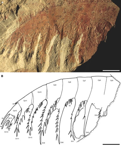
Figure 12. Echidnacaris briggsi (Nedin, Citation1995) comb. nov. frontal appendages. A, B, SAMA P49151b. C, D, SAMA P48371a. Scale bars: A = 20 mm; B, D = 5 mm; C = 10 mm.
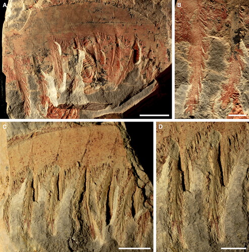
Figure 13. Echidnacaris briggsi (Nedin, Citation1995) comb. nov. head elements. A, SAMA P51380a. B, C, SAMA P45911b, a, respectively (light in C is from the top). Arrowheads in A and C indicate narrow marginal rim. Scale bars: 10 mm.
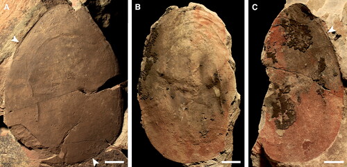
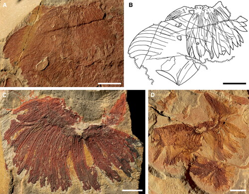
Figure 14. Echidnacaris briggsi (Nedin, Citation1995) comb. nov. oral cones. A, B, SAMA P57418. Photograph and camera lucida drawing, respectively. C, SAMA P55646a. D, SAMA P48195. Scale bars: 10 mm.
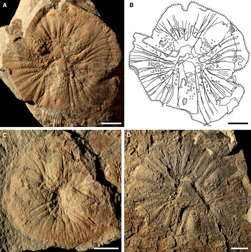
Figure 15. Echidnacaris briggsi (Nedin, Citation1995) comb. nov. oral cones. A, SAMA P57415a. B, SAMA P47415b. C, D, SAMA P52881a. C, overview. D, detail of teeth (arrowheads) at inner margin of a large plate (arrowhead in C). Scale bars: A–C = 10 mm; D = 1 mm.
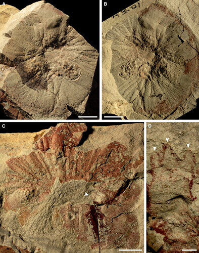
Figure 16. Echidnacaris briggsi (Nedin, Citation1995) comb. nov. oral cones. A, B, SAMA P55600. Deformed oral cone. A, SAMA P55600a. B, SAMA P55600b (light from upper right). C, SAMA P55650a. Detail of teeth (arrowheads) on inner margin of large and medium-sized plates. D, E, SAMA P55433. Smallest known oral cone. D, SAMA P55433a. E, SAMA P55433b. Scale bars: A, B = 10 mm; C = 5 mm; D, E = 2 mm.
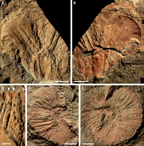
Figure 17. Unassigned Emu Bay Shale radiodont body flaps. A, SAMA P50326a. Largest known body flap. B, SAMA P55654b. C, SAMA P57407a. Flap with straight margin of articulation (arrowhead). D, SAMA P48154a, flap with 14 transverse lines and sharply defined margin of attachment (arrowhead). Scale bars: A = 20 mm; B–D = 10 mm.
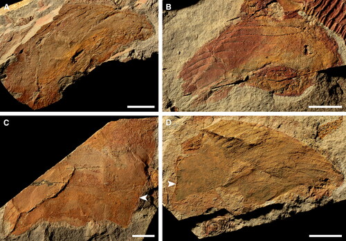
Figure 18. Unassigned Emu Bay Shale radiodont setal blades. A, B, SAMA P54822. Body flap and setal blades. A, SAMA P54822a. B, camera lucida drawing incorporating information from counterpart SAMA P54822b. C, SAMA P50287. D, SAMA P43611a. Scale bars: A, B, D = 10 mm; C = 5 mm.