Figures & data
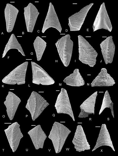
Figure 1. Comparative morphology of thoracican cirripedes. A, balanomorph Chionelasmus darwini (Pilsbry, Citation1907) in lateral view; B, C, verrucid Altiverruca quadrangularis (Hoek, Citation1883), lateral views; D, scalpellid Amigdaloscalpellum mamillatum (Aurivillius, Citation1898), lateral view. Abbreviations: FS, FT, fixed scuta and terga; MT, MS, moveable scuta and terga; ped, peduncular. Not to scale.
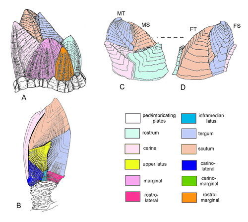
Figure 2. Comparative morphology of cirripedes. A, B, S, Pedupycnolepas articulata (Collins, Citation1980); A, cast of group of three specimens; B, reconstruction of peduncle; S, reconstruction. C, D, balanomorph Catophragmus pilsbryi Broch, Citation1922, in apical (C) and lateral (D) views. E, brachylepadid Parabrachylepas ifoensis (Withers, Citation1935), reconstruction in lateral view. F, brachylepadid Epibrachylepas newmani Gale, Citation2014b, reconstruction in lateral view. G–I, R, Etcheslepas durotrigensis Gale, Citation2014a, lateral views of articulated individuals (G–I); R, reconstruction. Note peduncle and presence of numerous lateral plates. J–L, verrucid Gibbosaverruca sp., note loss of peduncle and six-plated shell with strong asymmetrical differentiation of fixed and moveable scuta and terga (see also Fig. 3I–L). M, N, Q, shell of Brachylepas naissanti (Hébert, Citation1855) in lateral (M) and apical (N) views; Q, reconstruction, lateral view. Note large, hemiconical rostrum and carina and whorls of surrounding imbricating plates. O, Fallaxlepas fallax (Darwin, Citation1851a). P, Brachylepas guascoi (Bosquet, Citation1854). T, Pycnolepas rigida (J. de C. Sowerby, Citation1836), reconstruction in lateral view. U, Eoverruca hewitti Withers, Citation1935, reconstruction in lateral view. A, B, Aptian, Antarctic Peninsula; C, D, Recent, Panama; E, F, P, based on material from Campanian of Ivö Klack, Sweden; G–I, R, Tithonian, Kimmeridge Clay, Dorset, UK; J–L, Recent, Indian Ocean off Madagascar; M, N, O, upper Campanian, Norwich, Norfolk, UK; O, based on material from Maastrichtian, Rügen, Germany; T, based on material from Upper Albian, UK; U, based on material from Santonian, Suffolk, UK. Reconstructions (E, F, O–U not to scale. Colour key as in Figure 1. Abbreviations: c, carina; r, rostrum; rl, rostrolatus; s, scutum; ul, upper latus. Scale bars equal 5mm.
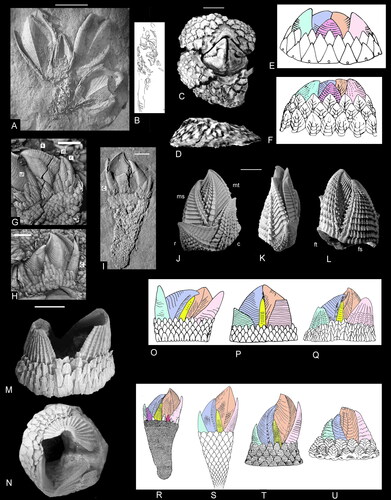
Figure 3. Morphology of upper latera and peduncular/imbricating plates. A–C, Etcheslepas durotrigensis Gale, Citation2014a; A, exterior view of upper latus; B, upper latus and other lateral plates on capitulum; C, peduncular plates. D, E, J–N, Pycnolepas rigida (J. de C. Sowerby, Citation1836); upper latus in D, external and E, internal views; J–N, peduncular plates. F, G, O, S, Brachylepas naissanti (Hébert, Citation1855), upper latus in F, external and G, internal views; O, external and S, internal views of imbricating plates. H, I, Q, R, Parabrachylepas ifoensis (Withers, Citation1935), upper latus in H, external and I, internal views; Q, R, imbricating plates. T–W, Eoverruca hewitti Withers, Citation1935; imbricating plates. P, Catophragmus pilsbryi Broch, Citation1922; interior view of imbricating plates. X, Brachylepas americana Zullo, Russell and Mellen, Citation1987, interior view of articulating imbricating plates. Scale bars equal 5 mm (B, D, E, P), 1 mm (A, C, X) and 0.5 mm (all others).
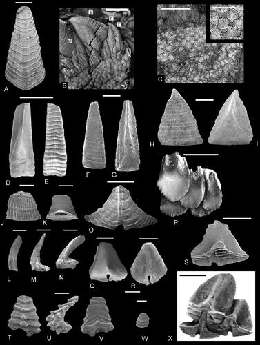
Figure 4. Terga and scuta of selected verrucomorph cirripedes, to show asymmetrical development and progressive differentiation of fixed and moveable valves. See text for discussion. A–D, Pycnolepas rigida (J. de C. Sowerby, Citation1836), A, B, terga and C, D, scuta. E–H, Eoverruca hewitti Withers, Citation1935, slight but distinct asymmetry of E, F, terga and G, H, scuta. I–L, Gibbosaverruca sp., strong asymmetry of I, J, terga and K, L, scuta. M–P, Verruca stroemia (O. F. Müller, Citation1776); note major differences in structure of fixed and moveable valves in M, N, terga and O, P, scuta. A–D, upper Albian, Gault Clay, Kent, UK. E–H, upper Santonian chalk, Suffolk, UK. I–L, Recent, Indian Ocean, off Madagascar. M–P, Recent, Donegal, Republic of Ireland. Abbreviations: abr, apicobasal ridge; ow, occludent wing. Scale bars equal 5 mm (A–D, I–L) and 0.5 mm (E–H, M–P).
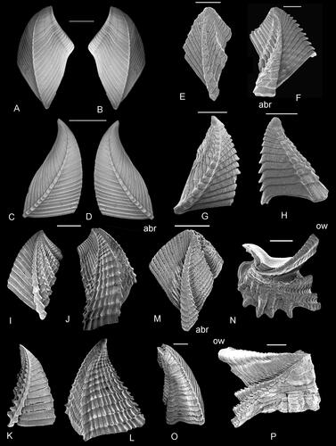
Figure 5. Nature of scutal–tergal articulations in thoracican cirripedes. A, Calantica sp. (Calanticomorpha); B, Capitulum sp. (Pollipedimorpha); C, Faxelepas bruennichi (Withers, Citation1914a) – Brachylepadomorpha, Pycnolepadidae); D, Brachylepas guascoi (Bosquet, Citation1854) – Brachylepadidae); E, Epibrachylepas newmani Gale, Citation2014b – Brachylepadomorpha, Brachylepadidae); F, Octomeris sulcata (Darwin, Citation1854) – Balanomorpha, Chthalamidae). Plesiomorphically (A, B), the scutum simply rests on the tergum, a weak tergal notch (tn, green) rests upon a poorly defined scutal auricle (sa, green). The upper latus infills the gap between scutum and tergum, and articulates with both (brown, x, y). In more derived forms (C, D, E), the scutal auricle and tergal notch become better defined and the articulation is more precise. In balanomorphs (F) the upper latus is lost, and the articulation between scutum and tergum is interpenetrant, and a prominent articular ridge (ar) on the scutum fits into an articular furrow (af) on the tergum. The scutal auricle and tergal notch are retained, and the regions of the scutum and tergum which previously articulated with the upper latus become a further articulation, the scutal wall (sw) and tergal wall (tw). Not to scale.
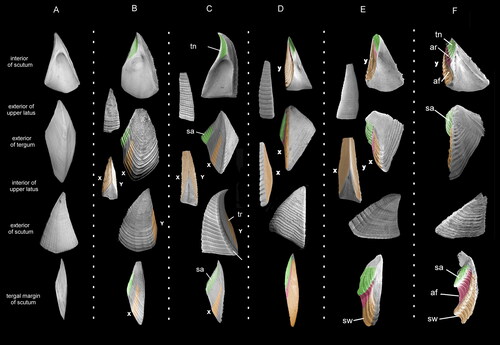
Figure 6. Comparative morphology of terga and scuta. A–C, pycnolepadid Pycnolepas rigida (J. de C. Sowerby, Citation1836), A, B, external and internal views of scutum; C, external view of tergum. F, G, H, M, N, brachylepadid Epibrachylepas newmani Gale in Gale & Sørensen, Citation2014; F, external, G, internal, H, tergal views of scuta, note presence of articular ridge (ar); M, N, external and internal views of terga; insertion sites for tergal depressor muscle indicated. D, E, O–Q, Balanomorph Octomeris brunnea (Darwin, Citation1854); D, external and E, tergal views of scutum, note articular ridge and furrow; O–P, tergum, in O, external, P, internal and Q, scutal views of tergum; note scutal auricle and articular furrow. I–L, brachylepadid Brachylepas guascoi (Bosquet, Citation1854); I, internal and J, external views of terga; K, external and L, internal views of scutum. Abbreviations: af, articular furrow; ar, articular ridge; sa, scutal auricle; td, tergal depressor muscle site; tn, tergal notch. Scale bars equal 5 mm (A–C, I–L), all others 1 mm.
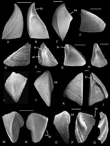
Figure 7. Morphological features of the rostrum and carina in brachylepadomorphs, verrucomorphs and balanomorphs. A–D, Faxelepas paronai (De Alessandri, Citation1895 – Pycnolepadidae); A, ventral view of rostrum; B, lateral view of rostrum; C, dorsal view of carina; D, interior view of carina. E, F, Etcheslepas durotrigensis Gale, Citation2014a; external views of E, rostrum and F, carina. G–J, Brachylepas naissanti (Hébert, Citation1855); G, ventral and H, internal views of rostrum; I, dorsal and J, internal views of carina. K–N, Catophragmus pilsbryi Broch, Citation1922 – Catophragmidae, Balanomorpha. K, ventral and L, internal views of rostrum; M, dorsal and N, internal views of carina. O–R, Eoverruca hewitti Withers, Citation1935 – Eoverrucidae; O, ventral and P, internal views of rostrum; Q, dorsal and R, internal views of carina. S, U, Gibbosaverruca sp. (Verrucidae); external views of S, rostrum and U, carina. T, V, Verruca stroemia (O. H. Müller, Citation1776); external views of T, rostrum and V, carina. Scale bars equal 10 mm (A–D), 5 mm (E–N, S–V) and 0.5 mm (O–R).
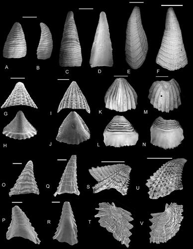
Figure 8. Comparative morphology of capitular plates of A, B, F, G, J–L, O, P, pollicipedid Etcheslepas fragilis (Withers, Citation1928) and C–E, H, I, M, N, Q, pycnolepadid Pycnolepas scalaris (Withers, Citation1914a). External views of A–D, terga, F, I, scuta, E, J, upper latera, K–N, carinae and O–Q, rostra. The overall structural and sculptural similarity of the valves can be viewed as evidence of close phylogenetic relationship. However, E. fragilis possesses numerous lateral plates, absent in P. scalaris. A, B, F, G, J–L, O, P, upper Kimmeridge Clay, Tithonian, Pavlovia pallasioides ammonite zone, Portland, Dorset, UK. C–E, H, I, M, N, Q, Grey Chalk subgroup, middle Cenomanian Turrilites acutus ammonite subzone, Dover, Kent, UK. Scale bars equal 0.5 mm (A, B, F, G, J, K, L, O, P), all others 0.2 mm.
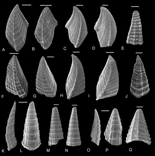
Figure 9. Stratigraphical distribution of taxa discussed in this paper.
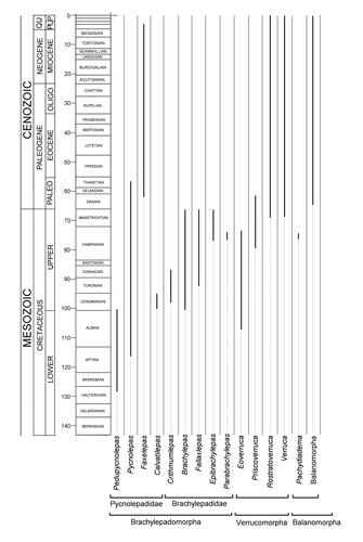
Figure 10. A, Cladogram, the most parsimonious tree (77 steps) from the equal weights analysis, based on analysis of 48 morphological characters () and 18 operational taxonomic units (OTUs), of which seven are extant taxa. Capitulum Gray, 1825 and Pollicipes Leach, Citation1817 were used as outgroups for the 16 in-group OTUs. One of the 48 characters was treated as continuous, using TNT (Goloboff & Catalano, Citation2016); all other characters were treated as unordered. Using the implicit enumeration tree search, both equal weights (EW) and implied weights analyses were implemented. Both analyses produced very similar single most parsimonious trees (MPTs). B, molecular tree based on mitogenome analysis, which shows Verrucomorpha as basal to all other Thoracicalcarea (after Gan et al., Citation2022, fig. 4). All other molecular and morphological studies place balanomorphs and verrucomorphs as sister groups. C, Thoracican cirripede classification and inferred phylogeny, based on nuclear genes and morphology, after Chan et al. (Citation2021, fig. 10). Note the close relationship of verrucomorphs and balanomorphs. Abbreviation: GER, GER, Gap Excess Ratio.
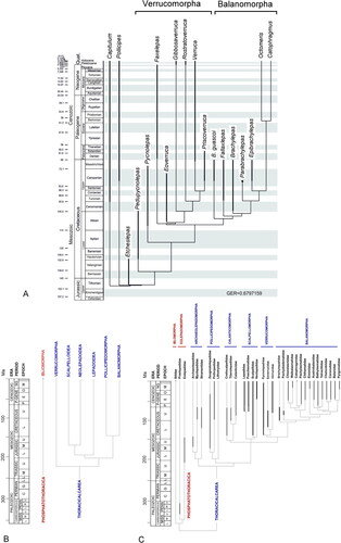
Table 1. Character list.
Table 2. Character scores.
Figure 11. Parallel evolution to sessile mode of life between the balanomorph-verrucomorph clade (left) and the neolepadoid clade (right). Verrucomorph-balanomorph clade; A, Etcheslepas durotrigensis Gale, Citation2014a; B, Pedupycnolepas articulata (Collins, Citation1980); C, Pycnolepas rigida (J. de C. Sowerby, Citation1836); D, Brachylepas naissanti (Hébert, Citation1855); E, Eoverruca hewitti Withers, Citation1935; F, Epibrachylepas newmani Gale, Citation2014b; G, Altiverruca quadrangularis (Hoek, Citation1883); H, Catomerus polymerus Darwin, Citation1854. Neolepadoid clade; I, Ashinkailepas seepiophila Yamaguchi, Newman and Hashimoto, Citation2004; J, Neoverruca brachylepadiformis Newman, Citation1969; K, Imbricaverruca yamaguchii Newman, Citation2000; L, Neobrachylepas relica Newman and Yamaguchi, Citation1995. Each lineage begins with pedunculate forms, and both subsequently give rise to asymmetrical forms with progressive differentiation of fixed and moveable valves (Verrucomorpha, Neoverrucidae) and symmetrical, low-profile forms with hemiconical carinae and rostra surrounded by imbricating plates (Balanomorpha, Neobrachylepadidae). Important differences between the two lineages include the distinctive paired processes on the apical interior of the scuta (Neolepadoidea, J–L) absent in the balanomorph-verrucomorph lineage. Additionally, molecular data consistently places the lineages in separate clades (see text). Abbreviations: FS, FT, fixed scuta and terga; MT, MS, moveable scuta and terga.
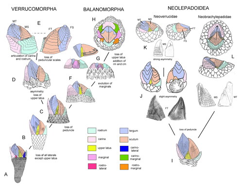
Figure 12. A–D, Pedupycnolepas lamellatus sp. nov. A, B, holotype scutum (NHMUK PI In 64815) in A, internal and B, external views. C, D, carinae (NHMUK PI In 64816, 64817) in dorsal view. E, F, Pedupycnolepas pulcher Gale, Citation2019, E, paratype rostrum, ventral view, original of Gale (Citation2019, fig. 11F; NHMUK IC 1398); F, holotype tergum in external view, original of Gale (Citation2019, fig. 11B; NHMUK IC 1397). G–Z, Calvatilepas recurvus gen. et sp. nov. G, H, V–Z, scuta (NHMUK PI In 64818, 64831–64834) in G, N, V–X, Z, external and H, Y, internal views. G, H, holotype (NHMUK PI In 64818); all other specimens are paratypes. I, J, peduncular plate (NHMUK PI In 64819) in I, external and J, internal views. K, M, O, Q, S, U, carinae (NHMUK PI In 64820, 64822, 64824, 64826, 64828, 64830) in K, O, S, U, dorsal, M, lateral and Q, internal views. P, R, T, rostra (NHMUK PI In 64825, 64827, 64829) in ventral view. L, Possible tergum, in external view (NHMUK PI In 64821). A–D, Grey Chalk Group, Cambridge Greensand Member, lower Cenomanian, Neostlingoceras carcitanense ammonite subzone, Barrington, Cambridge, UK. E, F, G–Z, Grey Chalk Group, Zig Zag Formation, upper Cenomanian Calycoceras guerangeri ammonite Zone, 70–72 m, Shakespeare Cliff, west of Dover, Kent, UK (Kennedy & Gale, Citation2006, fig. 2). E, F, lower Hauterivian Endemoceras amblygonium ammonite zone, Engelbostel, near Hannover, Germany. Scale bars equal 1 mm (F), 0.5 mm (E), 0.2 mm (A–D, G, H, K, L, N–V, X–Z) and 0.1 mm (M, W).
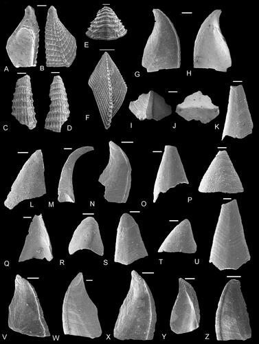
Figure 13. A–Q, Pycnolepas rígida (J. de C. Sowerby, Citation1836). A–C, scuta, in external (A, C) and internal (B) views (A, B, NHMUK PI In 64835, B, 64835a, C, 64835b). D–H, peduncular plates, originals of Gale (Citation2014b, fig. 4K–O; NHMUK In. 3224-8). I, tergum external view, original of Gale (Citation2014b, fig. 4I; NHMUK IC 1032). J, carina, dorsal view (NHMUK PI In 64837). K, L, Q, rostra; Q, ventral view, original of Gale (Citation2020a, fig. 4T; NHMUK IC 1035); K, L, external ventral (L) and internal (K) views (NHMUK PI In 64838). M–P, upper latera; M, N, original of Gale (Citation2014b, fig. 4R; NHMUK IC 1033). O, P (NHMUK PI In 64840) in external (N, P) and internal (M, O) views. R–Z, Pycnolepas batchelorum sp. nov. R, S, paratype upper latera (NHMUK PI In 64842, 64843) in internal (R) and external (S) views. T–V, terga (T, holotype, NHMUK PI In 64844, U, V, paratypes NHMUK PI In 64845, 64846) in external views. W, X, paratype scuta (NHMUK PI In 64847, 64848) in external views. Y, Z, paratype carinae (NHMUK Pi In 64849, 64850) in dorsal views. A, B, J, O, P, Gault Formation, upper Albian, Hysteroceras orbignyi ammonite Subzone, Ford Place, Wrotham, Kent, UK. I, M, N, Q, Hysteroceras varicosum ammonite subzone, Naccolt, near Wye, Kent, UK. C, middle Albian, Argiles à Tegulines, Lyelliceras lyelli ammonite zone, Aube, France. D–H, basal Gault Formation, lower Albian, 21 Acre Pit, Miletree Farm, Leighton Buzzard, Bedfordshire, UK. K, L, basal Gault Formation, lower Albian, Munday’s Hill, Leighton Buzzard, Bedfordshire, UK. R–Z, Bargate Formation, upper Aptian, Guildford, Surrey, UK. Scale bars equal; A–C, I–N, O–Q, 5 mm; D–H, 1 mm; R–Z, 0.3 mm.
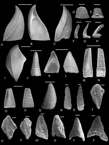
Figure 14. A–U, Pycnolepas scalaris (Withers, Citation1914a). A–C, external views of terga (NHMUK PI In 64851–64853). D, E, T, U, carinae (NHMUK PI In 64854, 64855, 64869) in D, E, U, external and T, internal views. F–H, J, scuta (NHMUK PI In 64856-64858, 64860) in F–H, external and J, internal views. I, K–P, rostra (NHMUK PI In 64862–64866) in I, K, L, N, P, ventral, M, lateral and O, internal views. Q–S, upper latera (NHMUK PI In 64867–64869) in Q, S, external and R, internal views. All from Grey Chalk Group, Zig Zag Formation, middle Cenomanian Turrilites acutus ammonite Subzone, 46.4 m (Kennedy & Gale, Citation2006, fig. 2), Samphire Hoe, west of Dover, Kent, UK. Scale bars equal 0.2 mm.
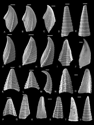
Figure 15. A–L, Faxelepas paronai (De Alessandri, Citation1895). A, B, upper latus, in A, exterior and B, interior views, original of Gale (Citation2020b, pl. 2, fig. 5; NHMUK IC 1826). C–F, scuta, in C, F, external and D, E, internal views. Originals of Gale (Citation2020b, pl. 2, figs 2–4; NHMUK IC 1823–1825). G, H, rostrum, in G, ventral and H, internal views (original of Gale, Citation2020b, pl. 2, fig. 11; NHMUK IC 1831). I, J, carina, in I, dorsal and J, internal views, original of Gale (Citation2020b, pl. 2, fig. 9; NHMUK IC 1829). K, external views of tergum, original of Gale (Citation2020a, pl. 2, fig. 6; NHMUK IC 1827). L–W, Faxelepas bruennichi (Withers, Citation1914a). M, N, upper latus, in M, external and N, internal views (NHMUK PI In 64871). O, carina, in dorsal view (NHMUK PI In 64872). P, carina in dorsal view, original of Gale (Citation2014b, fig. 4A; NHMUK IC 1022). Q, ventral view of rostrum (NHMUK PI In 64873). L, R, S, scuta, in L, S, internal and R, external views (NHMUK PI In 64875–64877). T, tergum in external view (NHMUK Pi In 64878). U, V, W, X, imbricating plates, in V, W, external, X, lateral and U, internal views (NHMUK PI In 64879–64881). Y-A2, Faxelepas landenica (Withers, Citation1953). Y, Z, external and internal views of scutum (NHMUK In. 38043). A1, Rostrum, ventral view (NHMUK In. 38046). A2, tergum, external view (NHMUK In. 38045). A–K, Plio-Pleistocene, Rodrigues Ridge, Indian Ocean. L–X, middle Danian, Faxe, Denmark. Y–A2, Selandian, Heers Formation, Wansin, north-east of Namur, Belgium. Scale bars equal 10 mm (C–L), 5 mm (A, B, M–S, Y–A1) and 0.4 mm (T–V, W, X).
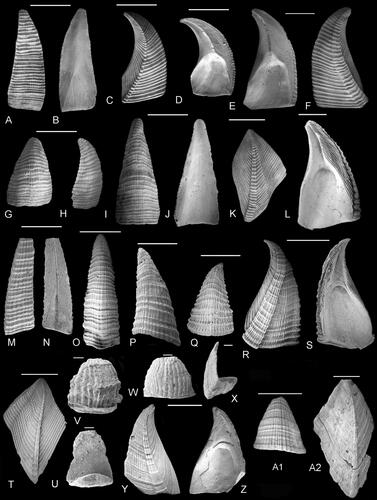
Figure 16. A–X, Eoverruca hewitti Withers, Citation1935. A–F, calcified basis, incorporating imbricating plates, in A, E, F, basal, B, apical and C, D, lateral views, originals of Gale Citation2014b, fig. 17A–F (NHMUK IC 1066). G–N, imbricating plates, in H, I–K, M, N, external and G, L, internal views, originals of Gale (Citation2014b, fig. 18A–H: NHMUK IC 1070–1076). O, P, fixed scutum in O, internal and P, external views, original of Gale (Citation2014b, fig. 19K, L: NHMUK IC 1061). Q–U, imbricating plates, originals of Gale (Citation2014b, fig. 17G–K: NHMUK IC 1060, 1067, 1068). V, moveable scutum, original of Gale (Citation2014b, fig. 19J: NHMUK IC 1065). W, moveable tergum, original of Gale (Citation2014b, fig. 19A: NHMUK IC 1063). X, fixed tergum, external view (NHMUK IC 1064). Upper Santonian, Uintacrinus socialis Zone, Hinderclay Lane, Wattisfield, Suffolk, UK. Scale bars equal 0.5 mm.
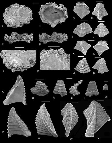
Figure 17. Distribution of Eoverruca hewitti in the upper Santonian Uintacrinus socialis zone across Europe, from Biocieniec, near Warsaw, Poland (5) through the Anglo-Paris Basin (1–4). The species occurs commonly within the lower part of the range of the zonal crinoid.
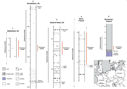
Figure 18. A, B, Eoverruca aubensis Gale, Citation2020a, holotype scutum in A, internal and B, external views, original of Gale (Citation2020a, fig. 14I, J; NHMUK IC 1566). C–K, Eoverruca hewitti Withers, Citation1935. C, Small moveable scutum, original of Gale (Citation2014b, fig. 19M: NHMUK IC 1089). D, F–H, carinae, in D, internal, F, H, dorsal and G, lateral views, originals of Gale, (Citation2014b, fig. 18I–L: NHMUK IC 1057, 1077, 1078). E, I–K, rostra, in E, internal, I, K, ventral and J, lateral views, originals of Gale (Citation2014b, fig. 18M–P: NHMUK IC 1058, 1080-1082). L–A4, Eoverruca symmetrica Gale, Citation2020a, paratypes. L–N, calcified basis incorporating imbricating plates, original of Gale (Citation2020a, fig. 14A–C: NHMUK IC 1552). O, Q, R, T–V, external views of terga, originals of Gale (Citation2020a, fig. 15, A, C, D, F–H: NHMUK IC 1557, 1559, 1561, 1563–1565). P, S, X, Y, scuta, in P, internal and S, X, Y, external views, originals of Gale (Citation2020a, fig. 15B, E, I, J: NHMUK IC 1558, 1562, 1566, 1567). Z–A4, carinae or rostra, in Z, A4, external, A1, A3, internal and A2, apical views, originals of Gale (Citation2020a, fig. 15L–P: NHMUK IC 1570–1573). A5, Eoverruca barringtonensis sp. nov. holotype, moveable tergum, external view (NHMUK PI In 64882). A, B, middle Albian, Anahoplites intermedius ammonite Subzone, Pogains, Aube, France. C–K, upper Santonian, Uintacrinus socialis Zone, Hinderclay Lane, Wattisfield, Suffolk, UK. L–A4, upper Campanian, Belemnitella woodi belemnite zone, uppermost Weybourne Chalk, Catton Grove, Catton, Norwich, Norfolk, UK. A5, Cambridge Greensand, lower Cenomanian, Neostlingoceras carcitanense ammonite Subzone, Barrington, Cambridgeshire, UK. Scale bars all equal 0.5 mm.
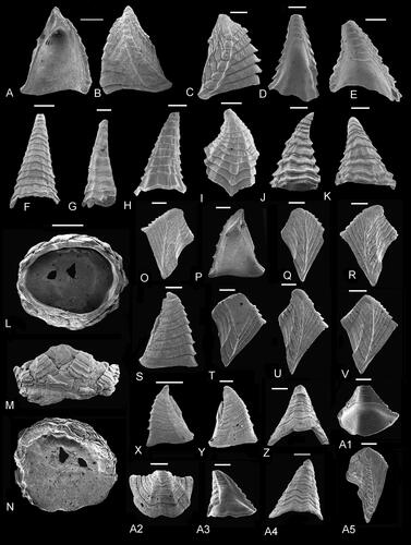
Figure 19. A–C, F–H, Youngiverruca ruegenensis Gale, Citation2014b. A–C, paratype fixed scutum, in A, external, B, internal and C, oblique views, original of Gale (Citation2014b, fig. 20J–L: NHMUK I. 6222), original of Withers (Citation1923, pl. 2, figs 46a, b). F–H, holotype fixed tergum in F, external, G, lateral and H, internal views, original of Gale (Citation2014b, fig. 20F–H: Withers Citation1923, pl. 2, fig. 48; Withers, Citation1935, pl. 45, fig. 13: NHMUK In. 16224). D, E, I–K, Priscoverruca elongata Gale, Citation2014b; D, E, paratype fixed scutum, in D, lateral and E, internal views, original of Gale (Citation2014b, fig. 22D, E: NHMUK 16225); I–K, holotype fixed tergum in I, external, J, lateral and K, internal views, original of Gale (Citation2014b, fig. 22A–C: NHMUK 16224). L, M, Rostratoverruca baxteri Gale, Citation2020c, holotype fixed scutum in L, external and M, internal views, original of Gale (Citation2020c, pl. 14, figs 1a, b: NHMUK IC 1768). N, O, Rostratoverruca romettensis (Seguenza, Citation1873); N, apical view of shell, original of Gale et al. (Citation2021, pl. 5, fig. 7b: PMC I. I. R. CIR-45); O, fixed tergum, external view, original of Gale et al. (Citation2021, pl. 5, fig. 11: PMC R. I. Cal. CIR-49). P, Rostratoverruca nexa (Darwin, Citation1854), figured after Darwin (Citation1854, pl. 21, fig. 5). Q, Rostratoverruca pusilla (Bosquet, Citation1857), figured after Bosquet (Citation1857, pl. 1, fig. 3). Specimens not found. Withers (Citation1935) selected the rostrum (B) as lectotype. A–K, lower Maastrictian, Rügen, Germany. L, M, Plio–Pleistocene, Rodrigues Ridge, Indian Ocean. N, Recent, Mediterranean, off Sicily, Italy. O, Pliocene, Scoppo, near Messina, Sicily, Italy. P, Recent, West Indies. Q, Maastrichtian, locality uncertain, localities listed for species are St. Pietersburg, Guelhem, and between Vilt and Sibbe, Limburg, the Netherlands. Abbreviations: C, carina; FT, fixed tergum; FS, fixed scutum; MS, moveable scutum; MT, moveable tergum; R, rostrum. Scale bars equal 0.5 mm (A–C, F–H, L–O), 1 mm (D, E, I–K) and 2 mm (P, Q).
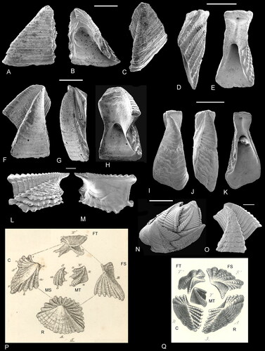
Figure 20. A–Y, Priscoverruca prisca (Bosquet, Citation1854). C–E, H–J, fixed scuta, in A–E, internal and H–J, external views (NHMUK PI In 64883–64887). F, G, K, moveable scuta, in F, G, external and K, internal views (NHMUK PI In 64888–64890). L–O, Q, fixed terga, in L, M, external, O, lateral and N, Q, internal views (NHMUK PI In 64891, 64892). P, moveable tergum, external view (NHMUK PI In 64893). R, rostrum, oblique apical view (NHMUK PI In 64894). S, T, carina in S, external and T, internal views (NHMUK PI In 64895). U, apical view of complete shell, original of Withers (Citation1913, fig. 2a, b; Withers, Citation1935, p. 342, figs 41, 42; Gale, Citation2014b, fig. 3F: NHMUK In. 27156). V, interior, X, apical and Y, lateral views of complete shell, original of Jagt and Collins (Citation1989, fig. 4d: NHMUK In. 62170). W, interior view of entire shell, original of Jagt and Collins (Citation1989, fig. 4: NHMUK In. 62171). A–T, upper Campanian, lower Belemnitella mucronata belemnite zone, Cringleford, Norwich, Norfolk, UK. U, upper Campanian, Belemnitella mucronata Zone, pit 154, Whitlingham, Norwich, Norfolk, UK. V–Y, upper lower Maastrichtian, Haccourt, Belgium. Scale bars all equal 0.5 mm.
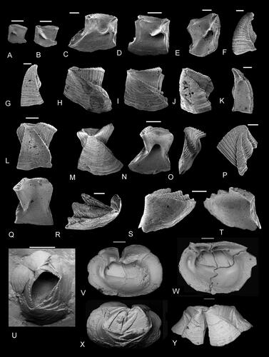
Figure 21. A–F, Bosquet’s type material of Verruca prisca Bosquet, Citation1854. A, B, fixed scutum, A, external, B, internal views, original of Bosquet (Citation1854, pl. 1, fig. 1a, b). C, moveable tergum, original of Bosquet (Citation1854, pl. 1, fig. 3a). D, moveable tergum, not figured by Bosquet. E, F, fixed tergum, E, internal and F, external views, original of Bosquet (Citation1854, pl. 1, fig. 5). Material in the Brussels Natural History Museum, all from Maastrichtian of St. Pietersberg, Maastricht, the Netherlands. G, Reproduction of the upper part of Bosquet (Citation1854, pl. 1). Scale bars equal 0.5 mm.
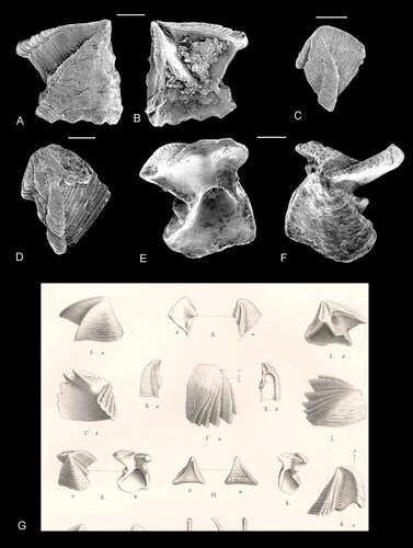
Figure 22. A–D, Verruca jagti Gale, Citation2014b. Holotype (NHMM JJ 13472), original of Gale (Citation2014b, fig. 23A). A complete individual in B, internal, D, apical and C, lateral views, with enlargement of moveable valves (A). E–M, Verruca stroemia (O. F. Müller, Citation1776), for comparison with fossil species. E, external view of moveable tergum; F, G, external and internal views of moveable scutum; H, internal view of shell; I, lateral view of shell; J, apical view of shell; K, oblique view of fixed tergum; L, M, fixed scutum, in L, internal and M, external views. A–D, from the ENCI-Heidelberg Cement Group quarry, Maastricht, southern Limburg, the Netherlands; Meerssen Member (Maastricht Formation, latest Maastrichtian), top 10 cm of subunit IVf-1 (hardground surface). E–M, Recent, Murvagh beach, County Donegal, Republic of Ireland. Scale bars equal 1 mm (B–D, H–I) and 0.5 mm (A, E–G, K–M).
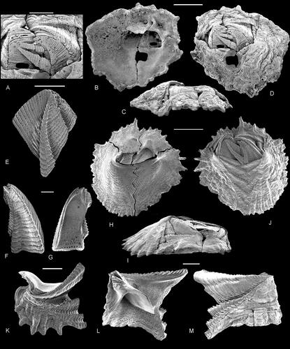
Figure 23. A–S, Brachylepas naissanti (Hébert, Citation1855). A, lateral and B, apical views of capitulum, original of Woodward (Citation1901, pl. 8, fig. 4; Withers, Citation1935, pl. 49, fig. 1a, b; Gale & Sørensen, Citation2014, fig. 3A–D: NHMUK In. 27160). C, D, G, J, upper latera, C, J, external and D, G, internal views (NHMUK PI In 64896, 64900, 64901). E, F, H, I, carinae, in E, H, dorsal and F, I, internal views (NHMUK PI In 64897, 64898). K, tergum, external view (NHMUK PI In 64902). L, M, rostrum, in L, ventral and M, internal views (NHMUK PI In 64899). N, O, scutum, in N, external and O, internal views (NHMUK PI In 64903). P–S, imbricating plates, in P–R, external and S, internal views (NHMUK PI In 64904–64907). A, B, White Chalk Subgroup, Belemnitella mucronata belemnite zone, upper Campanian, pit 153, Thorpe, Norwich, UK. C–S, White Chalk Subgroup, lower Belemnitella mucronata belemnite zone, upper Campanian, Cringleford, Norwich, UK. Scale bars equal 5 mm (A, B), all others 0.5 mm.
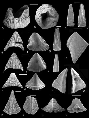
Figure 24. A–D, Fallaxlepas nervosa (Alekseev, Citation2009); A, tergum, original of Alekseev (Citation2009, pl. 4, fig. 20A); B, C, carina, in B, dorsal and C, lateral views, original of Alekseev (Citation2009, fig. 16A, B); D, rostrum in ventral view, original of Alekseev (Citation2009, fig. 15A). E, F, Brachylepas hantonensis Gale, Citation2020a. Holotype carina, original of Gale (Citation2020a, fig. 16C, D) in E, apical and F, lateral views (NHMUK IC 1575). G, H, Brachylepas thieli Gale, Citation2020a, holotype carina, original of Gale (Citation2020a, fig. 16A, B) in G, apical and H, lateral views (NHMUK IC 1574). I–Z, Brachylepas guascoi (Bosquet, Citation1857). I, J, rostrum in I, internal and J, ventral views, original of Gale and Sørensen (Citation2014, fig. 15E, F: NHMUK IC 842). K–M, carina, in K, dorsal, L, lateral and M, internal views, original of Gale in Gale and Sørensen (Citation2014, fig. 15A–C: NHMUK IC 841). N, O, apical views of small carinae, originals of Gale in Gale and Sørensen (Citation2014, fig. 14O, P: NHMUK IC 839, 840). P, Q, scutum in P, external and Q, internal views, original of Gale in Gale and Sørensen (Citation2014, fig. 14A, B: NHMUK IC 827). R, tergal view of scutum, original of Gale in Gale and Sørensen (Citation2014, fig. 14E: NHMUK IC 823). S, external view of tergum, original of Gale in Gale and Sørensen (Citation2014, fig. 14C: NHMUK IC 825). T–V, upper latera, in T, oblique lateral, U, external and V, internal views, originals of Gale in Gale and Sørensen (Citation2014, fig. 14I–K: NHMUK IC 826, 824, 836). W–A1, imbricating plates, in W, X, Z, external and Y, A1, internal views. Originals of Gale in Gale and Sørensen (Citation2014, fig. 15J, K: NHMUK IC 909, 910; NHMUK IC 913). A–D, Maastrichtian, Beshkosh Mountain, south-west Crimea, Ukraine. E, F, Lewes Chalk Formation, upper Turonian, Froxfield, pit no. 112 of Brydone, Citation1912, Hampshire, UK. G, H, lower Cenomanian, Kassenberg, Mülheim-Broich, Germany. I–A1, upper lower Campanian, Ivö Klack, Skåne, Sweden. Scale bars equal 5 mm (I–S), 1 mm (T–A1) and 0.5 mm (E–H).
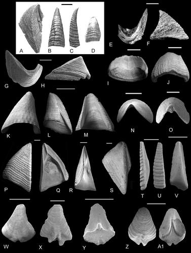
Figure 25. A–A1, Brachylepas americana Zullo, Russell and Mellen, Citation1987. A, B, carina, in A, dorsal and B, internal views (NHMUK In. 62268a). C, scutum, view of tergal surface (NHMUK In. 62268b). D–F, scutum in C, tergal, D, external and E, internal views (NHMUK In. 62268c). G, H, carina, in G, dorsal and H, internal views (NHMUK In. 62268d). I, J, scutum, in J, external and K, internal views (NHMUK In. 62268e). K, L, carina, in K, dorsal and L, lateral views (NHMUK In. 62268f). M–O, external views of terga (NHMUK In. 62268g-i). P–R, X, Y, imbricating plates, in P, R, Y, external and Q, X, internal views (NHMUK In. 62268g–k). Z, A1, upper latera, in Z, external and A1, internal views (NHMUK In. 62268l, m). S–W, rostra, in S, V, ventral, T, apical and U, W, internal views (S–U, In. 62268n; V, W, In. 62268o). A2–A5, Brachylepas angulosa Collins, Citation1973. Holotype carina (NHMUK In. 64479), original of Collins (Citation1973, pl. 3, fig. 16a–c) and Zullo et al. (Citation1987, figs 5, 8–10). A2, apical, A3, dorsal, A4, lateral and A5, internal views. A–Z, A1, basal Brownstown Formation, middle Campanian, Friendship, Hot Spring County, Arkansas, USA. A2–5, Ripley, Formation, Maastrichtian, Oktibbeha County, Mississippi, USA. Scale bars equal 5 mm (A–O, S–A5) and 0.5 mm (P–R, X–A1).

Figure 26. A–K, Fallaxlepas fallax (Darwin, Citation1851b). A, B, large scutum, A, external and B, internal aspects, original of Withers (Citation1935, pl. 48, fig. 2; NHMUK In. I. 14466). C, D, upper latus, C, external and D, internal views, original of Woodward (Citation1906, fig. 21) and Withers (Citation1935, pl. 48, fig. 3; NHMUK In. 30121). E, tergum, external view (NHMUK I. 15766). F, Carina, dorsal view (NHMUK IC 1578). G, Associated valves, showing carina, rostrum and scutum, original of Gale (Citation2020a, fig. 16J; Kutscher collection, Munich). H, I, imbricating plate, H, external and I, internal views (NHMUK PI In 64908). J, K, imbricating plate, J, external and K, internal views (NHMUK PI In 64909). L, tergum, external view (Kutscher collection, Munich). M, large carina, dorsal view (NHMUK In. 30125). N, rostrum, ventral view (NHMUK In. 30126). O, scutum, external view (NHMUK In. 30017). P, tergum, external view (NHMUK In. 30015). Q, R, large rostrum, Q, external and R, internal views (NHMUK In. 30129). S, T, rostrum, S, internal and T, external views (NHMUK In. 30130). U, small carina, dorsal view (NHMUK PI In 64910). A, B, D, F, H–K, U, Upper Campanian, Belemnitella mucronata belemnite zone, Norwich, Norfolk, UK. G, L, lower Maastrichtian, Rügen, Germany. E, Maastrictian, Benzenrathof, near Heerlen, the Netherlands. C, D, M–T, lower Maastrichtian, Belemnella sumensis belemnite zone, Trimingham, Norfolk, UK. Abbreviations: c, carina; r, rostrum; sc, scutum. Scale bars equal 5 mm (A–G, L–T) and 1 mm (H–K, U).
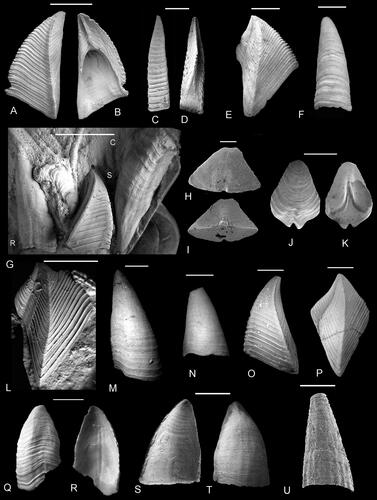
Figure 27. A–V, Parabrachylepas ifoensis (Withers, Citation1935). A, B, G, H, scuta, in A, H, external and B, G, internal views, originals of Gale in Gale and Sørensen (Citation2014, fig. 18A–D: NHMUK IC 881, 883). C, D, upper latus, in C, external and D, internal views, originals of Gale in Gale and Sørensen (Citation2014, fig. 18N, O: NHMUK IC 890). E, F, tergum, in E, internal and F, external views, originals of Gale in Gale and Sørensen (Citation2014, fig. 18F, G: NHMUK IC 855). I, J, K, marginal plates in I, K, external and J, internal views, originals of Gale in Gale and Sørensen (Citation2014, fig. 18I, J, K: NHMUK IC 887, 889). N, O, S, U, rostra, in N, dorsal, O, internal, U, lateral and S, apical views, originals of Gale in Gale and Sørensen (Citation2014, fig. 19B, C, E, G: NHMUK IC 893, 894, 895, 896). T, carina in apical view, originals of Gale in Gale and Sørensen (Citation2014, fig. 19A: NHMUK IC 892). L, M, P–R, V, Imbricating plates in M, P, R, external and L, Q, V, internal views, originals of Gale in Gale and Sørensen (Citation2014, fig. 19N–R: NHMUK IC 902–906). All from upper lower Campanian, Ivö Klack, Skåne, Sweden. Scale bars equal 1 mm (A–H, I–K, N, O, S–U) and 0.5 mm (L, M, P–R, V).
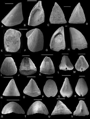
Figure 28. A–U, Epibrachylepas newmani Gale, in Gale and Sørensen, Citation2014. A–E, paratype scuta, in external (A–C), tergal (D) and internal (E) views, originals of Gale, in Gale and Sørensen, Citation2014, fig. 16A–D (NHMUK IC 855-857, 825); fig. 17I (NHMUK IC 876) E, original of Gale in Gale and Sørensen (Citation2014, fig. 17I: NHMUK IC 876). F–H, terga, in F, G, external and H, internal views; F, holotype, original of Gale in Gale and Sørensen (Citation2014, fig. 16E: NHMUK IC 858); G, H, paratypes, originals of Gale in Gale and Sørensen (Citation2014, fig. 16F, H: NHMUK IC 859, 861). I, J, marginal plate in I, external and J, internal views, original of Gale in Gale and Sørensen (Citation2014, fig. 17A, B: NHMUK IC 869). K, L, upper latera, in K, external and L, internal views, originals of Gale in Gale and Sørensen (Citation2014, fig. 16I–K: NHMUK IC 830, 831). O, P, rostra, in P, internal and O, apical views, originals of Gale in Gale and Sørensen (Citation2014, fig. 16M, N: NHMUK IC 864, 865). T, U, carinae, in T, apical and U, internal views, originals of Gale in Gale and Sørensen (Citation2014, fig. 16L, O: NHMUK IC 863, 866). M, N, Q–S, imbricating plates, in N, Q, R, S, external and M, internal views, originals of Gale in Gale and Sørensen (Citation2014, fig. 17C, G, H, J–L: NHMUK IC 870, 874, 875, 877–9). V, W, Epibrachylepas smeetsi (Bosquet, Citation1857) lectotype scutum, figured after Bosquet (Citation1857, pl. 3, fig. 11a–c), specimen presumed lost. A–U from upper lower Campanian, Ivö Klack, Skåne, Sweden. V, W, Maastrichtian, between Vilt and Sibbe, Limburg, the Netherlands. Scale bars equal 1 mm.
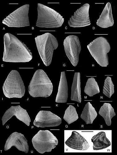
Figure 29. A–N, Crithmumlepas aycliffensis gen. et sp. nov. A, B, G, H, J, terga in external view (NHMUK PI In 64911, 64912, 64915, 64916, 64918); A is holotype (NHMUK PI In 64911), all other figured valves are paratypes. C–E, carina, in dorsal (C), lateral (D) and internal (E) views (NHMUK PI In 64913). I, K, L, scuta, in external (I, K) and internal (L) views (NHMUK PI In 64917, 64919). F, M, N, rostra, in ventral (F, N) and internal (M) views (NHMUK PI In 64914, 64920). O–X, Crithmumlepas hoensis gen et sp. nov. O, P, T–V, terga, in external views (NHMUK PI In 64921, 64922, 64925, 64927); V (NHMUK PI In 64927) is holotype all other figured valves are paratypes. Q–S, X, carinae, in dorsal (Q, S, X) and lateral (R) views (NHMUK PI In 64923, 64924, 64929). W, scutum, in external view (NHMUK PI In 64928). I–F, H–J, Grey Chalk Group, Zig Zag Formation, upper Cenomanian Calycoceras guerangeri ammonite Zone, 70–72m (Kennedy and Gale, Citation2006, fig. 2), Shakespeare Cliff, west of Dover, Kent, UK. O–X, Grey Chalk Group, Zig Zag Formation, middle Cenomanian Turrilites acutus ammonite Subzone, 46.4 m (Kennedy and Gale, Citation2006, fig. 2), Samphire Hoe, west of Dover, Kent, UK. G, White Chalk Group, Lewes Formation, Coniacian, upper Micraster cortestudinarium Zone, Hope Gap, Seaford, Sussex, UK. Scale bars equal; C–E, 0.3 mm, all others 0.2 mm.