Figures & data
Figure 1. Map of China showing the localities for Tianyusaurus (Henan and Jianxi provinces) and Yechilacerta yingliangia gen. et sp. nov. (Jianxi Province), with an enlargement showing the two localities from which the holotype of Yechilacerta yingiangia, YLSNHM01796 (site 1), and the second specimen, YLSNHM01791 (site 2), were recovered.
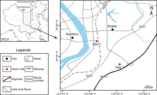
Figure 2. The holotype skull of Yechilacerta yingliangia gen. et sp. nov., YLSNHM01796, in (A) dorsal, (B) ventral, (C) right lateral, and (D) left lateral views. Abbreviations: An, angular; Ar, articular; Bo, basioccipital; D, dentary; Ect, ectopterygoid; Fr, frontal; ipv, interpterygoid vacuity; Ju, jugal; Mx, maxilla; N, nasal; Oco, occipital condyle; Pa, parietal; Pal, palatine; PBs, parabasisphenoid; Pf, postfrontal; Po, postorbital; ppr, paroccipital process; Prf, prefrontal; Pt, pterygoid; Pt.fs, pterygoideus fossa; Qu, quadrate; Sp, splenial; Sq, squamosal; Sur, surangular; V, vomer.
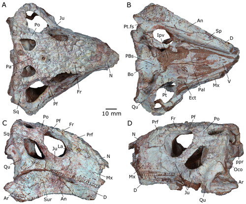
Figure 3. The holotype skull of Yechilacerta yingliangia gen. et sp. nov., YLSNHM01796, in (A) frontal, (B) occipital, and (C) oblique occipital views. Abbreviations: An, angular; b.tb, basal tubera; Bo, basioccipital; D, dentary; Ex, exoccipital; f.v, fenestra vestibuli; Ju, jugal; l.a.c, lateral adductor chamber; lrst, lateral opening of the recessus scalae tympani; Md, mandible; Mx, maxilla; N, nasal; Or, orbit; Pa, parietal; Pa.f, parietal foramen; ppr, paroccipital process; Pt, pterygoid; Qu, quadrate; sh, shelf; SMx, septomaxilla; So, supraoccipital; Sq, squamosal; Sur, surangular; v.f, vagus foramen.
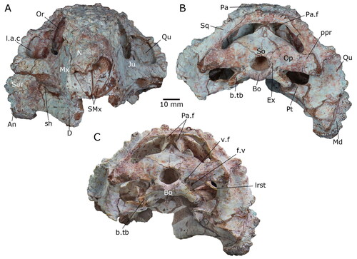
Figure 4. The skull of the second specimen of Yechilacerta yingiangia gen et sp. nov., YLSNHM01791, in (A) frontal, (B) dorsal, (C) right lateral, and (D) left lateral views.
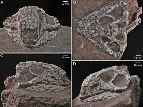
Figure 5. The holotype skull of Yechilacerta yingliangia gen. et sp. nov., YLSNHM01796 (A) in oblique ventral view to show the medial aspect of the right mandible, and (B) in ventral view to show the detached dentary symphysis and a fragment of premaxillary dentition. Abbreviations: Add.fs, adductor fossa; An, angular; D, dentary; Pr.a, prearticular; Sp, splenial.
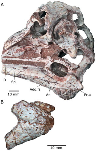
Figure 6. The holotype skull of Yechilacerta yingliangia gen. et sp. nov., YLSNHM01796. (A) and (B), right maxillary dentition with (A) anterior region and (B) posterior section, and (C) left maxillary and dentary dentition.
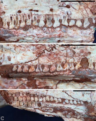
Figure 7. The postcranial skeleton of the second specimen of Yechilacerta yingiangia gen et sp. nov., YLSNHM01791 in dorsal view. A, pectoral region, forelimbs and anterior vertebral column; and B, dorsal, sacral and anterior caudal vertebrae, pelvic girdle, and hind limbs. Abbreviations: CV5, fifth cervical vertebra; Dig, digit (probably digit 4); ?DV1, interpreted position of first dorsal vertebra; Fe, femur; H, humerus; H.e, humeral epiphysis; Ili., ilium; Pu, pubis; Ra, radius; R.M, right manus; Sc, scapula; Ti, tibia; Ul, ulna.
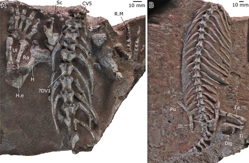
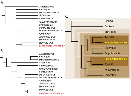
Figure 8. A and B, alternative consensus trees from a constrained TNT analysis of the emended Gauthier et al. (Citation2012) data matrix, with different levels of resolution. C, a summary tree of squamate relationships, based on molecular data, with the two most common alternative positions of Polyglyphanodontia reported by previous authors and the current paper. See text for further details.