Figures & data
Figure 1. Professor D.J.H. Cockayne FRS.

Figure 2. Professor A.F. Moodie, Dr P. Goodman, and Professor J.M. Cowley.
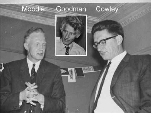
Figure 3. Comparison of experimental and calculated intensities as a function of incident angle for the 100 reflection in MoS2 Citation2. Reprinted with permission from Review of Scientific Instruments, D.J.H. Cockayne, P. Goodman, J.C. Mills and A.F. Moodie, Design and operation of an electron diffraction camera for the study of small crystalline regions, Vol. 38, Issue 8, p. 1097, Copyright 1967, American Institute of Physics.
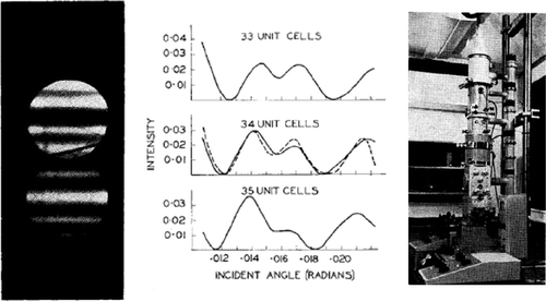
Figure 4. David Cockayne in 1967 in the laboratory photograph of the Department of Metallurgy, University of Oxford.
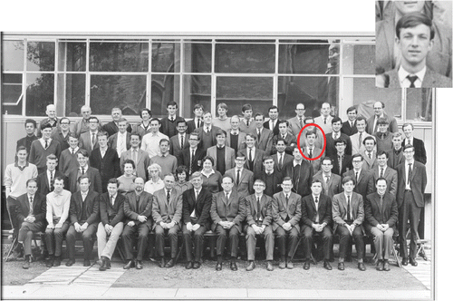
Figure 5. Metallurgy Annex at 10 Parks Road. Electron microscopes were housed in the hut.
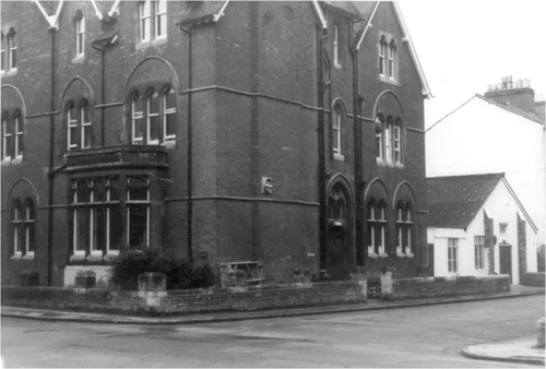
Figure 6. Climbing dislocation in Si imaged in dark-field (a) at the reflecting position and (b) in the weak beam condition Citation5. Reprinted from I.L.F. Ray and D.J.H. Cockayne, Proceedings of the Royal Society A, Vol. 325, p. 543, , 1971, with permission from The Royal Society.
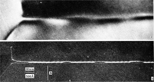
Figure 7. RDF analysis of thin film amorphous carbon. See text for details. Image created by D.J.H. Cockayne.
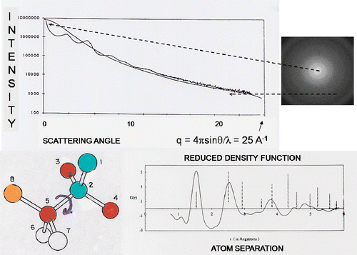
Figure 8. Structure of the C70 molecule from RDF data Citation11. See text for details. Reprinted by permission from Macmillan Publishers Ltd.: Nature, The structure of the C70 molecule, D.R. McKenzie, C.A. Davies, D.J.H. Cockayne, D.A. Muller and A.M. Vassallo, Vol. 355, p. 622, Copyright 1992.
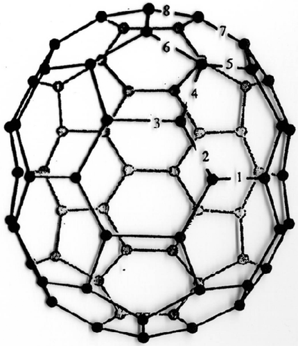
Figure 9. Comparison of experimental and simulated [001] zone axis images of InGaAs quantum dots on a GaAs substrate, indicating surface segregation of In. See text for details. Image created by D.J.H. Cockayne.
![Figure 9. Comparison of experimental and simulated [001] zone axis images of InGaAs quantum dots on a GaAs substrate, indicating surface segregation of In. See text for details. Image created by D.J.H. Cockayne.](/cms/asset/92fb7090-3a2d-41d8-a398-2bcb1bd5b1ad/tphm_a_439909_o_f0009g.gif)
Figure 10. High-angle annular dark-field STEM images of La and Lu doping atoms in intergranular glassy film between Si3N4 grains. See text for details. Image created by D.J.H. Cockayne.
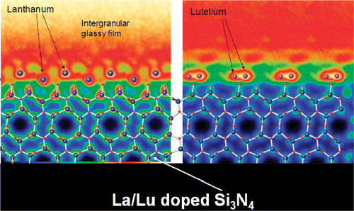
Figure 11. Microscopes on the move! Image created by D.J.H. Cockayne.
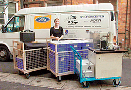
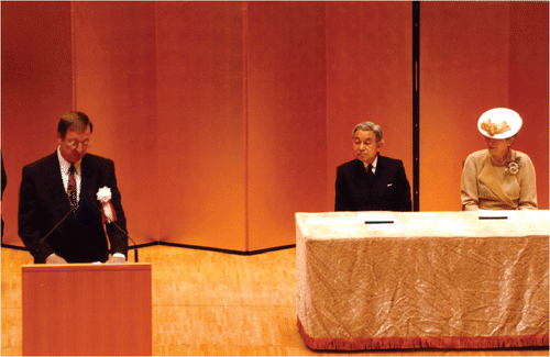
Figure 12. Cockayne delivering the opening address at the 16th International Microscopy Congress, Sapporo 2006, in the presence of the Emperor and Empress of Japan.