Figures & data
Figure 1. (colour online) Schematics of the location of the (a) tetrahedral (TIS) and (b) octahedral interstitial sites (OIS) for possible carbon occupancy in -Fe; (c) the classification of the OIS into ‘a’, ‘b’ and ‘c’ sites, and (d) representation of the so-called
-Fe
C
supercell structure, which accounts for the ordered product of spinodal decomposition, adapted from [Citation5].
![Figure 1. (colour online) Schematics of the location of the (a) tetrahedral (TIS) and (b) octahedral interstitial sites (OIS) for possible carbon occupancy in -Fe; (c) the classification of the OIS into ‘a’, ‘b’ and ‘c’ sites, and (d) representation of the so-called -FeC supercell structure, which accounts for the ordered product of spinodal decomposition, adapted from [Citation5].](/cms/asset/3715be6b-15bc-4620-9a9a-916f36582d55/tphm_a_1211790_f0001_oc.gif)
Figure 2. Illustration of the fundamental differences in the thermodynamics of (a) spinodal decomposition and (b) carbon segregation to defects. denotes the bcc phase without defects and miscibility gap.
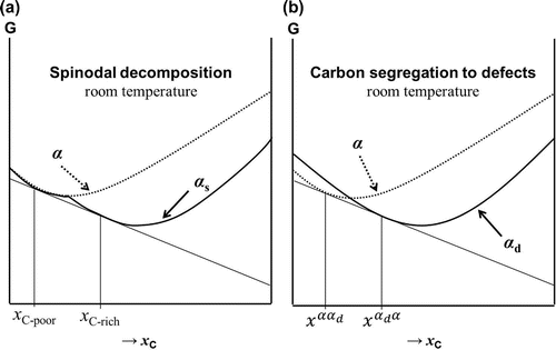
Table 1. Chemical composition of alloys studied.
Figure 3. Schematic representation of the thermal cycles for aged (A) and aged & tempered (A&T) heat treatments.
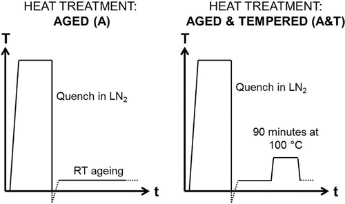
Figure 4. (colour online) Scans showing 20–50 2
, showing all labelled ferrite and austenite peaks where LC and HC refer to Fe–25Ni–0.19C and Fe–10Ni–0.87C wt.% alloys, and (a) shows the XRD patterns for samples that have undergone two weeks of room temperature ageing (A), and (b) shows XRD patterns after one week of room temperature ageing followed by tempering at 100
C for 90 min (A&T).
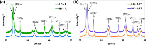
Table 2. Summary of XRD Rietveld refinement analysis, showing the global carbon content in solid solution based on matrix tetragonality.
Figure 5. (colour online) XRD scans shown for 13–37 2
where
,
and
carbides peak positions have been marked, for (a) Fe–25Ni–0.19C (LC) and (b) Fe–10Ni–0.87C wt.% (HC). The closed symbols represent the strongest reflections for each carbide.
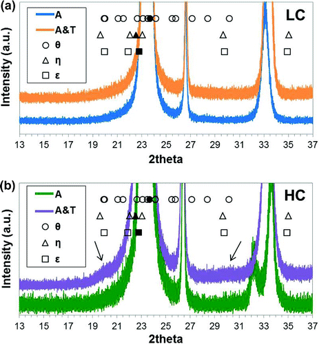
Table 3. Expected peak position for superlattice FeC
.
Figure 6. (colour online) Carbon maps of the reconstructed tips for steel LC (Fe–25Ni–0.19C wt.%) aged for five weeks showing (a) side view and (b) top view, and after 10 weeks of ageing showing (c) side view and (d) top view. Measurements were carried out under voltage pulse mode at 50 K using a 200 kHz pulse rate and 20 % pulse fraction. The arrows indicate visible carbon enrichment patterns in the reconstruction.
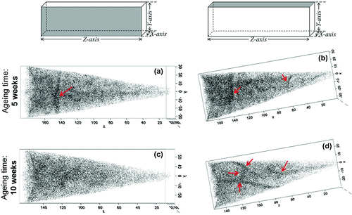
Figure 7. (colour online) Carbon maps of the reconstructed tips for steel LC (Fe–25Ni–0.19C wt.%) aged for five weeks showing (a) side view and (b) top view, and after 10 weeks of ageing showing (c) side view and (d) top view. Measurements were carried out under laser mode at 20 K using a 30 pJ laser energy and a 200 kHz pulse rate. The arrows indicate visible carbon enrichment patterns in the reconstruction.
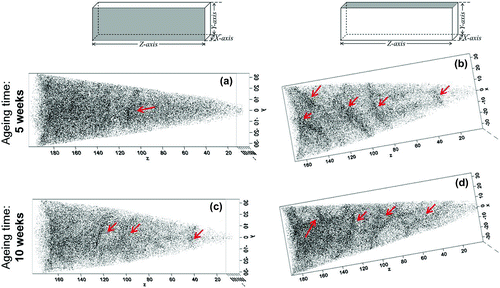
Figure 8. (colour online) Quantitative carbon analysis in Fe–25Ni–0.19 wt.% after 10 weeks of ageing at room temperature.
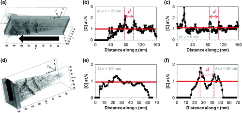
Figure 9. Schematic representation of the precipitation of carbides, in (a) cubic , and (b)
.