
Figures & data
Figure 1. Variation of free energy with the size of a precipitate when nucleating (a) homogeneously, (b) on a grain boundary and (c) on a dislocation.
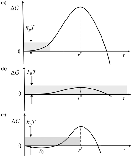
Figure 2. Time evolution of the number (i) of atoms in one cluster. “Reprinted (Fig. b) with permission from [Citation9] Copyright (2000) by the American Physical Society”.
![Figure 2. Time evolution of the number (i) of atoms in one cluster. “Reprinted (Fig. 7b) with permission from [Citation9] Copyright (2000) by the American Physical Society”.](/cms/asset/f0bcd3b4-827f-4c8d-8b5d-f54bffc0e221/tphm_a_1472400_f0002_b.gif)
Figure 3. Illustration of the exchange mechanism for interface controlled kinetics. Repetition of the same sequence will produce coarsening and reduction of the free energy.
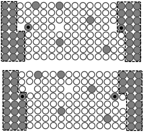
Figure 4. Solute concentration profile near the interface under different regimes.
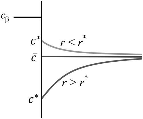
Figure 5. Evolution of a cluster size distribution from time t 0 to t 1. Adapted from [Citation23].
![Figure 5. Evolution of a cluster size distribution from time t 0 to t 1. Adapted from [Citation23].](/cms/asset/597211db-ab0a-43cd-9d84-b5fbf3d5491b/tphm_a_1472400_f0005_b.gif)
Figure 6. On the left: An isolated subcritical embryo experiencing a net loss of atoms. On the right: A cloud of subcritical embryos.
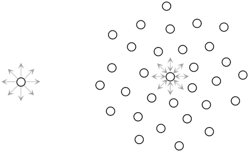
Figure 7. Left: A cloud containing a high number density of embryos. Right: One of the dissolving embryos located at periphery.
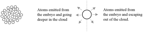
Figure 8. Plot of the factor F vs. time.
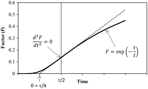
Figure 9. Evolution of the number density of embryos with respect of the radius of the embryos. Calculations were made with C 0 = 1 × 10²² m−3, r 0 = 2 nm and υ = 0.01 nm/s.
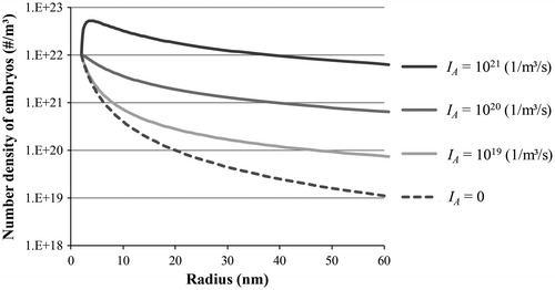
Table 1. Comparison between the classical nucleation theory and the subcritical growth theory (this contribution).
Figure 10. Schematized events captured by Liu et al. [Citation11] during the precipitation of θ′ in an Al-5.7 wt.%Cu specimen. Precipitates 1 and 3 are created and are then dissolved under the influence of precipitates 2 and 4. The boxes indicate the time in hours and minutes.
![Figure 10. Schematized events captured by Liu et al. [Citation11] during the precipitation of θ′ in an Al-5.7 wt.%Cu specimen. Precipitates 1 and 3 are created and are then dissolved under the influence of precipitates 2 and 4. The boxes indicate the time in hours and minutes.](/cms/asset/e86a2501-0de4-41fe-91bb-db6733203eab/tphm_a_1472400_f0010_b.gif)