Figures & data
TABLE 1. Summary of Clinical Information and Pathologic Diagnoses Based on ConventionalHistology and Immunohistochemistry
Figure 1. Very early hydropic abortion at 10 × lens (A and B) and 20 × lens (C and D) hematoxylin and eosin stain (A and C) and positive staining for p57 (B and D) in the cytotrophoblast, mesenchymal cells, and intermediate trophoblast.
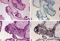
Figure 2. Partial mole at 10 × lens (A and B) and 20 × lens (C and D) show the hematoxylin and eosin stain (A and C) and focal positivity for p57 (B and D) in the cytotrophoblast but not the mesenchyme. The intermediate trophoblast is focally positive.
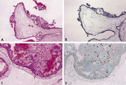
Figure 3. Complete mole is seen at 10 × lens (A and B) and 20 × lens (C and D) hematoxylin and eosin stain (A and C). There is no p57 staining (B and D) in the cytotrophoblast or in the intermediate trophoblast.
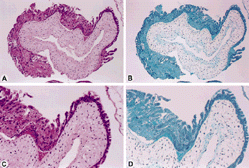
TABLE 2. Diagnosis and Clinical Outcome
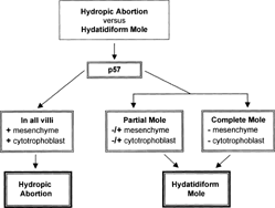
Figure 4. Recommended diagnostic approach to hydropic abortions and hydatidiform moles.