Figures & data
Table 1. Sampling sites, collection dates and plant species collected.
Figure 1. (a) The San Bernardino site (1930 m a.s.l.) during snowmelt on 8 June 2014; arrowheads indicate places where felty brown to black mycelial mats of Allantophomospsis cytisporea as shown in (b), (c), and (d) can be observed. (b) Mycelial mat right at the edge of melting snow. (c) Mycelial mat covering horsts of dead leaves of the grass Nardus stricta. (d) Mycelial mat covering Calluna vulgaris. (e) Shoot blight on C. vulgaris observed at the San Bernardino site on 24 August 2014. (f) Close-up of (e).
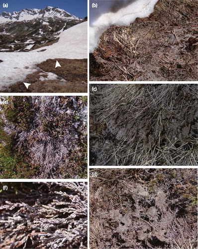
Table 2. Allantophomopsis strains isolated from mycelial mats covering shoots of various plant species at various sites in the Swiss Alps during snow melt.
Table 3. Collection details and GenBank accession numbers of isolates included in the phylogenetic analysis.
Figure 2. (a) Mycelium of Allantophomopsis cytisporea colonizing dead tissue of a Calluna vulgaris leaf (scale bar: 20 µm); the bright meandering lines are the cell walls of the puzzle piece–like epidermal cells of C. vulgaris. (b) Interaction between Herpotrichia pinetorum (strain 95-Fi; Schneider et al. Citation2009) to the left and A. cytisporea (strain A_DA_21_15) to the right after 21 days at 4°C on MEA. Conidia of A. cytisporea strains (c) A_SB_8_12 and (d) A_SB_6_12 in H2O stained with fountain pen ink (Pelikan, blue) and observed using phase contrast optics (scale bar: 10 µm); arrowheads indicate the slimy appendages of the conidia.
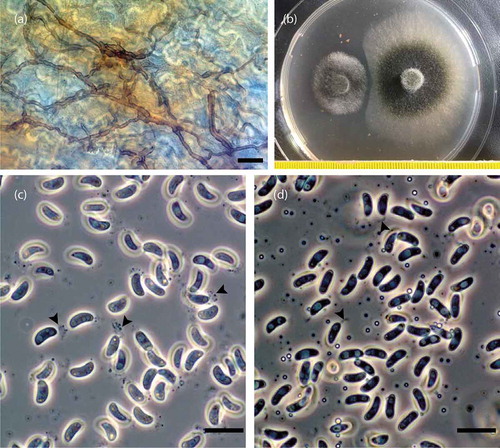
Figure 3. The most parsimonious tree showing the phylogenetic relationships among snow mold strains collected for this study (bold) and related species as inferred from ITS1-5.8s-ITS2 (489 character states including gaps) sequences. The scale bar shows the number of changes, and bootstrap support values of greater than 50 percent from 100 replicates are shown at the nodes. Taxon names are preceeded by GenBank accession numbers. The species boundaries are delimited with colored blocks. The tree was rooted to Gremmenia (Phacidium) infestans (GenBank accession U92305).
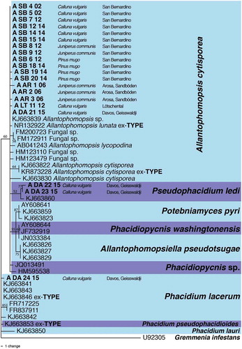
Table 4. Frequency (percentage) of incubated mycelial samples giving rise to Allantophomopsis cytisporea, Herpotrichia pinetorum, and other fungi.
Figure 4. Growth rates of two Allantophomopsis cytisporea strains and Herpotrichia pinetorum at various temperatures.