Figures & data
Table 1. Effect of low-folate culture conditions on the expression of DNMT1, DNMT3A and DNMT3B mRNA expression in the colon cancer cell lines CaCo2 and WiDr.
Figure 1. DNMT1 expression in 8 cell lines from various histological sources normalized to β-actin expression. RaijB is a Burkits lymphoma, RT112 and SW780 are bladder cancer, BT474 and C-33A are cervix carcinoma, LS174T is colon cancer, PANC-1 is pancreatic cancer and HEC-1A is endometrial cancer. The SEM in all assays was less <15%. No statistically significant differences were observed between cell lines with a different pathological origin.
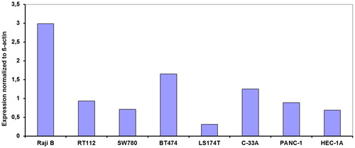
Table 2. Expression of DNMT1, DNMT3A and DNMT3B mRNA expression in 7 separate human cancer cell lines.
Figure 2. DNMT1 expression in 26 different xenografts including several patient derived xenografts (PDX) normalized to β-actin expression. DM77 is bone cancer, PANC-005, PANC-006, PANC-008, PANC-011, PANC-019, PANC-159, PANC-294, PANC-266, MD/JH-015, BxPC-3 are pancreatic cancer, DM75 is synovial cancer, PULM-009, BML-13, BML-6, H460 and H69 are lung cancer, Colo-205, HT29 and SW480 are colon cancer and MOLT-4, Daudi, H929, OPM-2 and WSU-DLCL are hematological malignancies. The SEM in all assays was less <15%. There was not statistically significant difference between the tumour types.
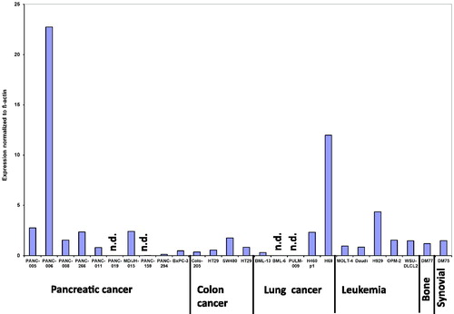
Table 3. Protein and mRNA expression levels of DNMT1, 3A and 3B in normal and corresponding tumour tissue
Figure 3. IHC staining of DNA methyltransferase 1, 3A and 3B protein in colorectal cancer (CRC) patients and DNMT1 in two 5 µm xenograft sections (G), selected for high and low DNMT1 mRNA expression. Positive immunoreactivity for DNMT1, 3A and 3B was found in CRC tissue (A, C, E) and corresponding normal tissue (B, D, F). Unlike DNMT3B that is present in the cytosol and nuclei (E insert), DNMT1 and DNMT3A are mainly well distributed only in the cytoplasm of tumour cells. DAB staining shows DNMT1 expression in brown. Pictures were taken with 40x lens magnification.
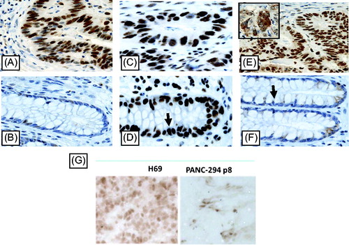
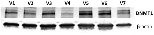
Figure 4. DNMT1 expression in white blood cells from 7 human volunteers (V1-V7) as determined with western blotting equal amounts of protein were used for each sample. β-actin was used as a loading control.