Figures & data
TABLE 1 Pregnancy outcome following fetal exposure to di-(2-ethylhexyl)phthalate for the last 16 days of gestationFootnote1
TABLE 2 Body weights and ano-genital distances (AGD) measured in offspring of Sprague-Dawley CD rats exposed to di- (2-ethylhexyl)phthalate for the last 16 days of gestation
FIG. 1 Nitrite (top) levels and tumor necrosis factor alpha (TNFα, bottom) produced by adherent splenocytes isolated from 5-week-old female offspring exposed to di-(2-ethylhexyl) phthalate for the last 16 days of gestation. Cells were stimulated with 10 ng/ml LPS and supernatants were collected after 24 hours of incubation at 37°C. The minimum detection limit for TNFα was 0.7 pg/ml. There were no statistical differences (p > 0.05).
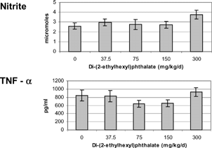
FIG. 2 Cytokines produced by unseparated splenocytes isolated from 5-week-old offspring exposed to di-(2-ethylhexyl)phthalate in utero for the last 16 days of gestation. Cells were incubated with 5 μg/ml Con A for 72 hours. The minimum detection limit for each of the cytokines was as follows: IFN-γ < 13 pg/ml, IL-2 < 5 pg/ml IL-4 < 1.3 pg/ml, IL-10 < 5 pg/ml, IL-12 < 3 pg/ml. There were no significant differences between treatments (p > 0.05).
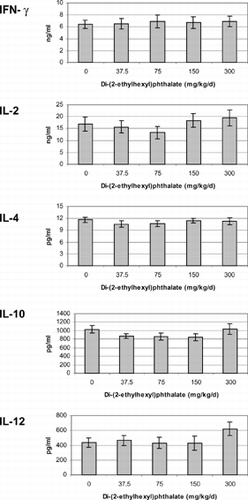
FIG. 3 Cytokines produced by unseparated splenocytes isolated from 13-week-old offspring exposed to di-(2-ethylhexyl)phthalate in utero for the last 16 days of gestation. Cells were incubated with 5 μg/ml Con A for 72 hours. The minimum detection limit for each of the cytokines was IFN-γ < 13 pg/ml, IL-2 < 5 pg/ml, IL-4 < 1.3 pg/ml, IL-10 < 5 pg/ml, IL-12 < 3 pg/ml. There were no significant differences between treatments (p > 0.05).
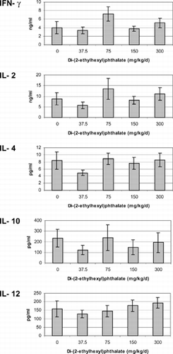
FIG. 4 Cytokines produced by unseparated splenocytes isolated from non-pregnant adults exposed to di-(2-ethylhexyl)phthalate for 16 days concurrently with pregnant dams. Cells were incubated with 5 μg/ml Con A for 72 hours. The minimum detection limit for each of the cytokines was as follows: Interferon gamma < 13 pg/ml, IL-2 < 5 pg/ml IL-4 < 1.3 pg/ml, IL-10 < 5 pg/ml, IL-12 < 3 pg/ml. There were no significant differences between treatments (p > 0.05).
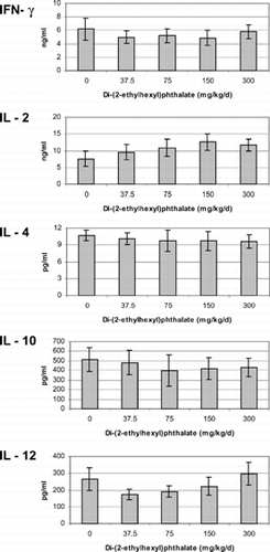
TABLE 3 Flow cytometry profile of cell populations in 5-week-old offspring exposed to di-(2-ethylhexyl)phthalate in utero for the last 16 days of gestation