Figures & data
Figure 1. Oscillating circuit of TheraCell device (Guth MediTec, Schechen, Germany (80 mT)).
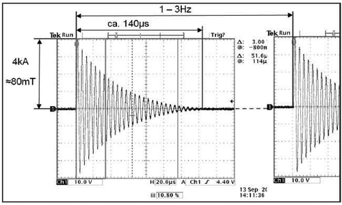
Figure 2. Treatment protocol.
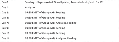
Figure 3. Placement of the 24-well plate by template in the center of the EMTT device loop.
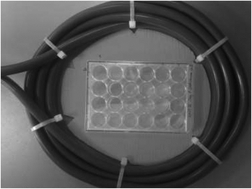
Figure 4. Primer list for real-time PCR.
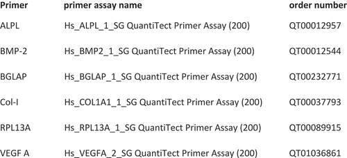
Figure 5. Group-specific MSC viability measured by MTS assay (n = 4; group A: 80 mT, group B: 150 mT, group C: control group). The optic absorption of all three groups increased significantly from day 1 to day 7 and day 14. *p < .05, n = 4; Tukey’s multiple comparison test; data are means ± SD.
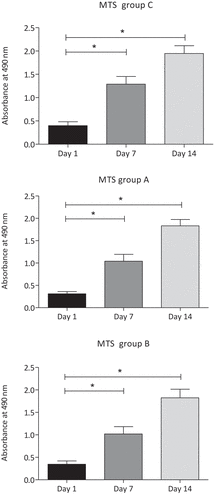
Figure 6. (a) and (b) Show relative cell viability measured by MTS assay of group A (80 mT) and group B (150 mT) compared with control group C (n = 4; group C = 100% cell viability) after 7 and 14 days of culture. No significant difference of optical density is measured between untreated group C and EMTT-stimulated groups A and B; Tukey’s multiple comparison test; data are means ± SD.
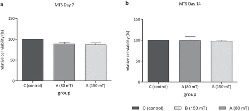
Figure 7. Relative expression of pro-osteogenic marker collagen I (Col I), alkaline phosphatase (ALPL), osteocalcin (BGLAP), vascular endothelial growth factor A (VEGF A) and bone morphogenic protein-2 (BMP-2) on day 7 (a) and day 14 (b) in group C (control group), group A (80 mT) and group B (150 mT) measured by real-time PCR. No significant difference of relative expression; n = 4, Tukey’s multiple comparison test.
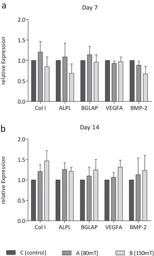
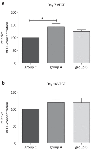
Figure 8. Relative VEGF concentration in group C (control), group A (80 mT EMTT) and group B (150 mT EMTT) on day 7 (a) and day 14 (B).*p < .05, n = 3; Tukey’s multiple comparison test.