
Figures & data
Figure 1. Experimental design used to study effects of di-n-octyltin dichloride (DOTC) on neurodevelopment of Wistar rats. Rats were exposed to DOTC at a dose of respectively 0, 3, 10 or 30 mg/kg of diet from two weeks before mating of F0-generation to postnatal day (PND) 61–70 of F1-generation, and a limited group of F1-animals up to PND 90** (conform OECD test guideline 443 for an extended one-generation reproductive toxicity study). Acclimatization to the housing conditions started at least a week prior to exposure*. During development, adolescence and adulthood physical, sensory and sexual landmarks were studied, a functional operational battery (FOB) and auditory startle response (ASR) were carried out, nociception and motor activity (MA) were tested, and effects on cognition were investigated performing passive and active avoidance tests (PA and AA, respectively). In addition, cerebrum and cerebellum were sampled for analysis by [18F]FDG PET functional imaging, magnetic resonance structural imaging (MRI), quantitative macroscopy (brain weight and specific gravity (SG)) and microarray gene expression profiling. Microarray gene expression profiling was applied to male offspring only (all dose groups); PET and MRI were limited to male F1-rats of the control and 30 mg/kg DOTC dose groups.
![Figure 1. Experimental design used to study effects of di-n-octyltin dichloride (DOTC) on neurodevelopment of Wistar rats. Rats were exposed to DOTC at a dose of respectively 0, 3, 10 or 30 mg/kg of diet from two weeks before mating of F0-generation to postnatal day (PND) 61–70 of F1-generation, and a limited group of F1-animals up to PND 90** (conform OECD test guideline 443 for an extended one-generation reproductive toxicity study). Acclimatization to the housing conditions started at least a week prior to exposure*. During development, adolescence and adulthood physical, sensory and sexual landmarks were studied, a functional operational battery (FOB) and auditory startle response (ASR) were carried out, nociception and motor activity (MA) were tested, and effects on cognition were investigated performing passive and active avoidance tests (PA and AA, respectively). In addition, cerebrum and cerebellum were sampled for analysis by [18F]FDG PET functional imaging, magnetic resonance structural imaging (MRI), quantitative macroscopy (brain weight and specific gravity (SG)) and microarray gene expression profiling. Microarray gene expression profiling was applied to male offspring only (all dose groups); PET and MRI were limited to male F1-rats of the control and 30 mg/kg DOTC dose groups.](/cms/asset/823e2f2d-8266-4f35-a06b-783625df31c4/itxm_a_2281610_f0001_c.jpg)
Table 1. Number of F1-animals in an extended one-generation reproductive toxicity study (OECD TG 443).
Figure 2. Braintree of Wistar rat, with relative change in volume data upon di-n-octyltin dichloride (DOTC) exposure, estimated from MR images taken on postnatal day (PND) 21 and 61. Insert gives an overview of the original ‘brain vesicles’ in the developing rat brain. The percentages presented per brain region (left: PND 21; right: PND 61) represent the effect of 30 mg/kg of diet DOTC on brain region volume, relative to the control group. Exposure occurred via the diet from 2 weeks pre-mating of F0 animals to PND 61–70 of the F1 generation. A practically overall enlargement of brain volume is seen at developmental/adolescence age (PND 21), which is no more observed at young adulthood (PND 61). Despite the fact that none of the enlargements observed at PND 61 differs significantly from the control group, the trajectories of brain development from early development to PND 21 and from PND 21 to PND 61 differ between control and DOTC groups. Statistical key: ANOVA with Dunnett’s post hoc comparison test: different from control, *p < .05 (thick red border lines); trend, # .05 < p < .1 (thin red border lines). OB: olfactory bulb; ATS: nucleus accumbens, olfactory tubercle, septum; PND: postnatal day.
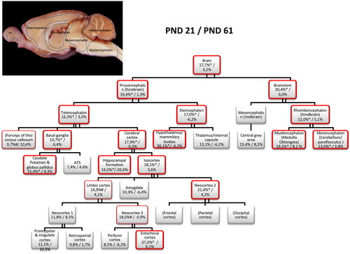
Figure 3. Motor activity of male rat offspring (N = 10/group) after exposure to di-n-octyltin dichloride (DOTC), 0 (control diet) or 30 mg/kg of diet DOTC from 2 weeks premating (F0-generation) up to postnatal day (PND) 61–70 (F1-generation). Motor activity was measured as the total distance moved (cm) during 30 min of motor activity testing on PND 13, 17, 21 and 61 ± 1. A tendency toward hyperactivity upon DOTC exposure is observed on PND 61 ± 1. Statistical key: ANOVA with Dunnett’s posthoc comparison: # .05 < p < .1.
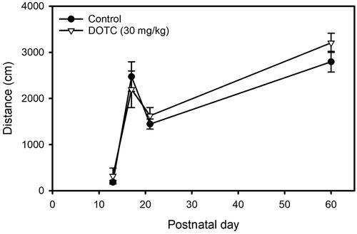
Figure 4. Auditory startle response (ASR) of male offspring (N = 10/group) after exposure to 0 (control) or 30 mg/kg of diet di-n-octyltin dichloride (DOTC) from 2 weeks premating (F0-generation) up to postnatal day (PND) 61–70 (F1-generation). The ASR was tested on PND 23 and expressed as the average voltage (V) per time-block. Hyper-responsiveness is observed at the start of testing. Statistical key: ANOVA with Dunnett’s posthoc comparison: * p < .05.
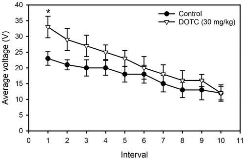
Table 2. Effect of exposure of male F1-rats to di-n-octyltin dichloride (30 mg/kg of diet) on brain volume, brain weight and specific gravity.
Table 3. Effect of exposure of male F1-rats to di-n-octyltin dichloride (30 mg/kg of diet) on volume of forebrain, midbrain and hindbrain regions estimated from magnetic resonance images using stereology (Cavalieri principle).
Figure 5. Brain volume (top panels; N = 4–6/group), brain weight (mid panels; N = 10/group) and specific gravity (bottom panels; N = 4–6/group) of male offspring after exposure to 0 (control) or 30 mg/kg of diet di-octyltin-di-chloride (DOTC) from 2 weeks premating (F0-generation) up to PND 61–70 (F1-generation). Brains are weighed at necropsy (postnatal day (PND) 21 or 69 ± 1) immediately after removal from the skull; brain (region) volumes are calculated from MRI scans taken in the living animal on PND 21 or 61, using stereology (Cavalieri principle). The weight of the brain represents the group mean of the sum weight of both hemispheres, whereas the weight of cerebrum and cerebellum represent the group mean of the weight in one hemisphere only (left/right chosen randomly). Statistical key: ANOVA with Dunnett’s post hoc comparison: * p < .05 different from control.
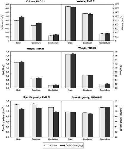
Table 4. Effect of exposure of male F1-rats to di-n-octyltin dichloride (30 mg/kg of diet) on volume of cerebral cortex (hippocampal formation and isocortex) subregions estimated from magnetic resonance images using stereology (Cavalieri principle).
Figure 6. [18F]FDG brain uptake (SUV) in nine regions of interest in male F1-rats (N = 3–5/group) upon exposure to 0 (control) or 30 mg/kg of diet di-n-octyltin dichloride (DOTC) from 2 weeks premating (F0-generation) up to PND 61–70 (F1-generation). [18F]FDG PET was performed on postnatal day (PND) 17, 21, 35, 61. The trajectories of brain development from birth to adolescence and from adolescence to adulthood differ between the two groups, but no between-group differences were observed at any timepoint (Kruskal–Wallis with Newman Keuls post-hoc comparison).
![Figure 6. [18F]FDG brain uptake (SUV) in nine regions of interest in male F1-rats (N = 3–5/group) upon exposure to 0 (control) or 30 mg/kg of diet di-n-octyltin dichloride (DOTC) from 2 weeks premating (F0-generation) up to PND 61–70 (F1-generation). [18F]FDG PET was performed on postnatal day (PND) 17, 21, 35, 61. The trajectories of brain development from birth to adolescence and from adolescence to adulthood differ between the two groups, but no between-group differences were observed at any timepoint (Kruskal–Wallis with Newman Keuls post-hoc comparison).](/cms/asset/39b334bf-aee6-4bab-afb1-f98873e20490/itxm_a_2281610_f0006_b.jpg)
Figure 7. Biological process categories significantly overrepresented among 52 differentially expressed genes upon di-n-octyltin dichloride (DOTC) treatment (male F1-animals; all doses and time points; p < .05 is at value of 1.3). exposure to DOTC at a dose of 0, 3, 10 or 30 mg/kg of diet occurred from 2 weeks premating (F0-generation) up to postnatal day 61–70 (F1-generation).
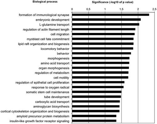
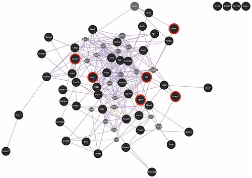
Figure 8. Network of 52 differentially expressed genes upon di-n-octyltin dichloride treatment (male F1-animals; all doses and time points). red circles mark genes involved in processes of neurodevelopment/neural function, and/or are associated with autism. Exposure to DOTC at a dose of 0, 3, 10 or 30 mg/kg of diet occurred from 2 weeks premating (F0-generation) up to postnatal day 61–70 (F1-generation).
Supplemental Material
Download MS Word (8.9 MB)Data availability statement
The data that support the findings of this study are available from the corresponding author, DMGdG, upon reasonable request.