Figures & data
Figure 1. Insulin priming effect on estradiol-induced growth of breast cancer MCF-7 cells. (A) Growth response of the insulin-primed and unprimed MCF-7 cells. Relative growth response was assessed using MTT assay as described in materials and methods. MCF-7 cells were treated with 1 nM of estradiol, 10 nM of insulin, 30 nM of IGF-1, 1 μM ICI 182,780 alone or co-treated with estradiol (top). Note that MCF-7-10-42 and MCF-7-1.7-48 represent independent batches of insulin-primed MCF-7 cells (middle). Both primed and unprimed MCF-7 cells were assayed for estradiol-induced growth response in a dose-dependent manner (2nd bottom) Both primed cells (3-day vs. 7-day priming) were assayed for estradiol-induced growth response in a dose-dependent manner (bottom). (B) The expression of ERα and insulin receptor (INSR) were identified using immunoblot assay after treatment of cells with 1 nM of estradiol and/or 10 nM of insulin for 48 h. (C) mRNA expression of PR in response to estradiol in both cell lines. Total RNAs were isolated from cells treated with 1 nM of estradiol alone or in combination with 1 μM of ICI182,780 for 24 h. (D) Functional evaluation of AR for growth response of the insulin primed and unprimed MCF-7 cells; protein expression of AR (Top left) and mRNA expression of an AR target FKBP51 upon DHT treatment (Top right). Growth response for DHT treatment was assessed using MTT assay after treatment with 1 nM DHT alone or in combination with 10 nM insulin or 1 μM bicalutamide (bottom). White bars-unprimed (□); black bars-primed cells (▪). Values are mean ± SEM. *P < 0.05; #P < 0.001.
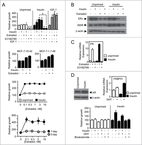
Figure 2. Estradiol-induced signaling depends on PI3K/Akt and ERK signaling. (A) Relative growth response of insulin-primed and unprimed MCF-7 cells. Cells were treated with 10 μM of MEK inhibitor U0126 or 20 μM of PI3K inhibitor LY294,002 in the presence or absence of 1 nM estradiol for 7 d and followed by MTT assay. (B) Immunoblot assay for protein expression in insulin signaling pathway. Total cell lysates were obtained from the cells treated with 1 nM of estradiol in a time-dependent manner and followed by further immunoblot assay for protein expression of interest. Densitometric analysis was performed. Values are mean ± SEM. #P < 0.001.
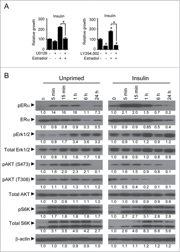
Figure 3. Growth response of the insulin-primed cells upon estradiol-mediated regulation of cell cycle factors. Immunoblot assay was performed for the expression of cell cycle factors involving cyclins (A, B, D1), cyclin-dependent kinase inhibitor (p21, p27) and apoptosis (Bcl-xL, BAX). Total cell lysates were obtained from both primed and unprimed cells after 24 h treatment with 1 nM estradiol alone or together with 1 μM of ICI182,780, 100 μM of LY294,002, 10 μM of U0126, or 3 mM of metformin. Densitometric analysis was performed.
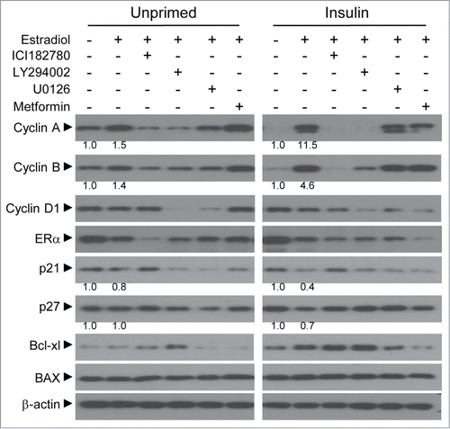
Figure 4. Estradiol modulation of gene expression involved in glucose metabolism. The QPCR assay was performed for metabolic genes in insulin-primed and unprimed cells. Total RNAs were isolated from cells treated with 1 nM of estradiol alone or in combination with 1 μM of ICI182,780 for 24 h. (A) Genes associated with glycolytic pathway. (B) Genes involved in serine biosynthesis from 3-phosphoglycerate. (C) Genes for pentose phosphate pathway for anaplerotic reaction to replenish glyceraldehyde-3-phosphate. (D) Schematic diagram of glycolysis and the associated biosynthetic pathways. Continuous bold arrows show proposed flow of carbon in insulin-primed cells after treatment with estradiol. White bars-unprimed (□); black bars-primed cells (▪). Values are mean ± SEM. *P < 0.05; ΔP < 0.01; #P < 0.001.
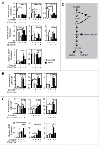
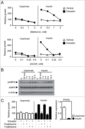
Figure 5. Therapeutic evaluation of anti-diabetic drugs in the insulin-primed breast cancer. (A) Growth response of both primed and unprimed breast cancer cells to metformin and AICAR. Cells were treated with metformin or AICAR in a dose-dependent manner in the presence or absence of 1 nM estradiol. (B) Immunoblot assay for phosphorylated-AMPK and total AMPK. Cells were treated with 5 mM of metformin in a time-dependent manner for immune blot assay of AMPK phosphorylation. (C) Growth response of both primed and unprimed breast cancer cells to TZDs. Cells were treated with 3 μM of pioglitazone or troglitazone in the presence or absence of 1 nM estradiol. The mRNA expression of PPAR γ were measured in both unprimed(□, cycle time = □24) and insulin primed (▪, cycle time = □28) MCF-7 cells.