Figures & data
Figure 1. MRI of IVC Tumor Thrombus in clear cell RCC before and after SABR. Coronal (top) and axial (bottom) contrast enhanced MR images at different time points during the course of treatment. After nephrectomy and thrombectomy, the patient had an intraluminal recurrence of tumor thrombus, which was adherent to the IVC wall (arrowheads, A). The superior extent of the thrombus is inferior to the diaphragm (Level III; arrow, A). Note the size of the thrombus at the level of the right hepatic vein (arrow, B). After systemic targeted therapy (C) there was obvious disease progression with thrombus extending superior to the diaphragm (level IV, arrow) and increased enhancement (arrowhead, C). Note marked increased in transverse diameter (arrow, D). Two years after SABR therapy there is persistent thrombus extending above the diaphragm (arrow, E) although exhibiting clear decrease in enhancement (arrowhead, E) and marked reduction in transverse diameter (arrow, F).
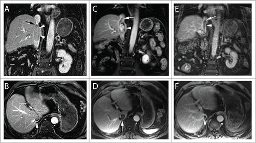
Figure 2. SABR Treatment of Recurrent IVC-TT. (A-C) Representative axial, sagittal, and coronal images of the SABR treatment plan with isodose lines showing dose distribution and coverage of the IVC-TT. Patient was immobilized with a vacuum bag in an Elekta body frame. Abdominal compression and 4D-CT with contrast was used for respiratory motion management and assessment respectively. Treatment planning MRI was fused for target delineation. The dose was prescribed to the 84% isodose line via 11 non-coplanar photon beams of 10 MV and 3D optimization ensuring >95% PTV coverage with a 0.5 cm margin on the TT. (D) Radiation dose volume histogram from SABR plan of 50 Gy in 5 fractions showing optimized doses to critical organs as well as target volume (PTV). (E) Radiation dose constraints used for treatment planning.
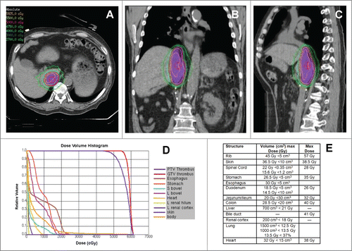
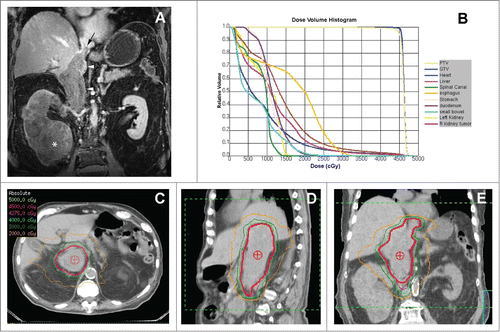
Figure 3. SABR Treatment of Unresectable IVC-TT. (A) Coronal contrast-enhanced MRI during the venous phase demonstrates low level enhancement in a large renal mass (asterisk), which infiltrates the entire right kidney parenchyma and extends superiorly with a expansile tumor thrombus in the inferior vena cava (arrowheads). Note the superior extent of the tumor thrombus (black arrow) above the diaphragm (i.e. level IV thrombus). (B) Radiation dose volume histogram from SABR plan of 45 Gy in 5 fractions showing optimized doses to critical organs and target volumes. The patient set-up and target delineation was similar to the first case. The plan required 13 non-coplanar photon beams of 10 MV and IMRT optimization to ensure >95% PTV coverage with a 0.5 cm margin on the TT. C-E) Representative axial, sagittal, and coronal images of the SABR treatment plan with isodose lines showing dose distribution and coverage of the IVC-TT.