2,897
Views
122
CrossRef citations to date
0
Altmetric
Research Papers

miRNA profiling in vitreous humor, vitreal exosomes and serum from uveal melanoma patients: Pathological and diagnostic implications

, , , , , , , , , , , , & show all
Pages 1387-1396 | Received 05 Jan 2015, Accepted 23 Apr 2015, Published online: 12 Aug 2015

Figures & data

Figure 1. Characterization of VH exosomes. (A) Average particle size in VH exosome samples was determined by dynamic light scattering. Y-axes: signal intensity (%); X-axes: size of particles (nm). (B) Flow cytometry detection of surface molecules on nanoparticles isolated from VH samples. The exosomes were bound to aldehyde-sulfate latex beads conjugated with anti-CD9, anti-CD63 or anti-CD81 antibodies and analyzed by flow cytometry. The antibodies were compared with their appropriate isotype control IgG1.
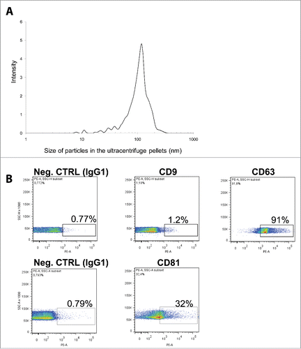
Figure 1. Characterization of VH exosomes. (A) Average particle size in VH exosome samples was determined by dynamic light scattering. Y-axes: signal intensity (%); X-axes: size of particles (nm). (B) Flow cytometry detection of surface molecules on nanoparticles isolated from VH samples. The exosomes were bound to aldehyde-sulfate latex beads conjugated with anti-CD9, anti-CD63 or anti-CD81 antibodies and analyzed by flow cytometry. The antibodies were compared with their appropriate isotype control IgG1.

Figure 2. Comparison of miRNAs found in VH, VH exosomes and serum of UM and healthy controls. (A) Venn diagrams showing the overlap between miRNA sets found in different types of samples. (B) Quantitative representation of miRNA different expression between UM patients and controls in VH, VH exosomes, serum. (C) Correlation between RQs from VH and its exosomes: x-axis represents the −log10 of RQ of vitreal miRNAs in UM patients with respect to normal controls; y-axis represents the −log10 of RQ of exosomal miRNAs in UM patients with respect to normal controls.
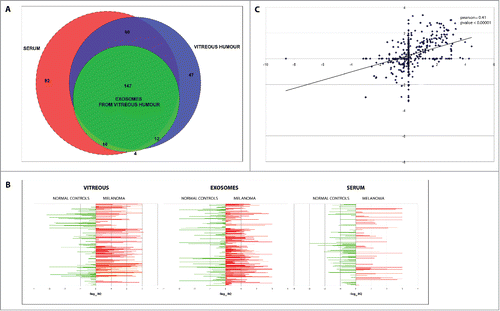
Figure 2. Comparison of miRNAs found in VH, VH exosomes and serum of UM and healthy controls. (A) Venn diagrams showing the overlap between miRNA sets found in different types of samples. (B) Quantitative representation of miRNA different expression between UM patients and controls in VH, VH exosomes, serum. (C) Correlation between RQs from VH and its exosomes: x-axis represents the −log10 of RQ of vitreal miRNAs in UM patients with respect to normal controls; y-axis represents the −log10 of RQ of exosomal miRNAs in UM patients with respect to normal controls.

Figure 3. Single TaqMan assays for miR-21, miR-34a, miR-146a, miR-618. Box plots representing the expression of: (A) miR-146a, (B) miR-21, (C) miR-34a, (D) miR-618, analyzed by single TaqMan assay on whole vitreous humor, exosomes from vitreous (ExoVitr.), whole serum, or exosomes from serum (ExoSer.) from an independent cohort of 12 patients. y-axis represents the –ΔCt of miRNAs in UM patients with respect to normal controls. Statistical significance was evaluated by the Wilcoxon rank sum test (p-value < 0.05).
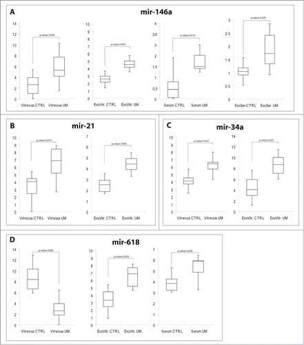
Figure 3. Single TaqMan assays for miR-21, miR-34a, miR-146a, miR-618. Box plots representing the expression of: (A) miR-146a, (B) miR-21, (C) miR-34a, (D) miR-618, analyzed by single TaqMan assay on whole vitreous humor, exosomes from vitreous (ExoVitr.), whole serum, or exosomes from serum (ExoSer.) from an independent cohort of 12 patients. y-axis represents the –ΔCt of miRNAs in UM patients with respect to normal controls. Statistical significance was evaluated by the Wilcoxon rank sum test (p-value < 0.05).
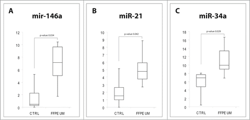
Table 2. Demographics, tumor parameters, time treatment/enucleation in UM

Table 1. DE miRNAs in UM patients

Figure 4. MiRNAs expression in FFPE UM specimens. Box plots representing the expression of: (A) miR-146a, (B) miR-21, (C) miR-34a, analyzed by single TaqMan assay on paraffin-embedded UM compared to healthy choroidal melanocytes. y-axis represents the −ΔCt of miRNAs in UM patients with respect to normal controls. Statistical significance was evaluated by the Wilcoxon rank sum test (p-value < 0.05).

Figure 4. MiRNAs expression in FFPE UM specimens. Box plots representing the expression of: (A) miR-146a, (B) miR-21, (C) miR-34a, analyzed by single TaqMan assay on paraffin-embedded UM compared to healthy choroidal melanocytes. y-axis represents the −ΔCt of miRNAs in UM patients with respect to normal controls. Statistical significance was evaluated by the Wilcoxon rank sum test (p-value < 0.05).
Supplemental material

Supplemental File 1

Download MS Excel (69.5 KB)

Reprints and Corporate Permissions

Please note: Selecting permissions does not provide access to the full text of the article, please see our help page How do I view content?

To request a reprint or corporate permissions for this article, please click on the relevant link below:

Academic Permissions

Please note: Selecting permissions does not provide access to the full text of the article, please see our help page How do I view content?

Obtain permissions instantly via Rightslink by clicking on the button below:

If you are unable to obtain permissions via Rightslink, please complete and submit this Permissions form. For more information, please visit our Permissions help page.