Figures & data
Table 1. Clinico-pathological association of 8p deletion
Figure 1. Examples of FISH findings using the 8p deletion probe. (A) Heterozygous deletion as indicated by the lack of one orange 8p signal. (B) Normal 8p copy numbers as indicated by 2 orange 8p signals and 2 green centromere 8 signals.
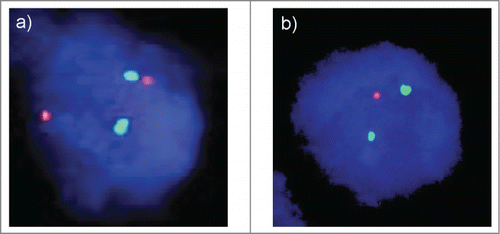
Figure 2. Associations between 8p status and amplifications of HER2, MYC, and CCND1 analyzed by FISH.
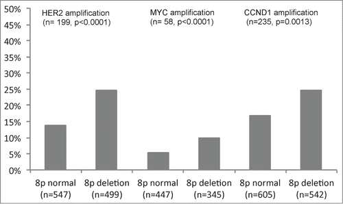
Figure 3. Association between 8p deletion and Ki67-labeling index. (A) all cancers, (B) Grade 1 cancers, (C) Grade 2 cancers, and (D) Grade 3 cancers.
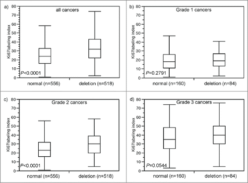
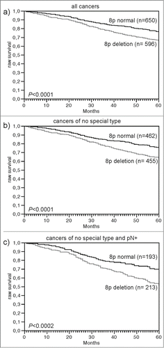
Figure 4. Association between 8p deletion and raw survival. (A) all cancers (n = 1,246), (B) no specific type cancers (n = 917), and (C) no specific type and pN positive cancers (n = 406).