Figures & data
Figure 1. IRF-1 selectively inhibits human breast cancer growth. (A) The spontaneously immortalized nonmalignant human breast cell line, MCF-10F, and the human breast cancer cell lines ZR75-1, MDA-MB-468, and the SK-BR-3 were infected with the control Ad-Ψ5 or Ad-IRF-1, at multiplicities of infection (MOIs) 25, 100, 10, and 10 respectively. Cell growth inhibition was measured by MTT assays as described in Material and Methods. (B) Cells were infected at varying MOIs of Ad-IRF-1 and cells were harvested 24 h later. Cellular lysates were prepared for immunoblotting as described in Material and Methods. IRF-1 expression was evaluated by immunoblotting in the MCF-10F, MDA-MB-468, and the SK-BR-3 cell lines. (C) The MCF-10F, MDA-MB-468, and SK-BR-3 cell lines were subsequently infected at MOIs of 50, 10, and 25 respectively. MTT assays were conducted and cell growth inhibition was calculated as described in Material and Methods. (D) HMEpC and MDA-MB-468 cells were infected at varying MOIs and cells were harvested for immunoblotting as described in Material and Methods. (E) HMEpC and MDA-MB-468 cells were infected at MOIs of 50 and 10 respectively. Cell growth inhibition assays were conducted as described in Material and Methods. MTT was added 48 h post infection.
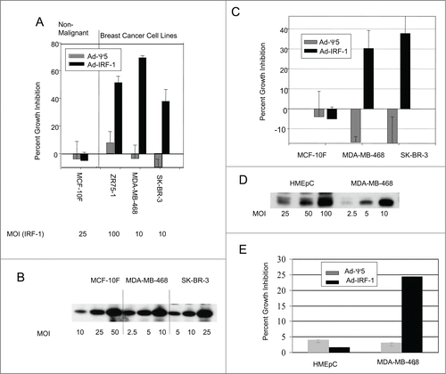
Figure 2. IRF-1 selectively induces apoptotic cell death in human breast cancer cell lines. (A) The nonmalignant human mammary epithelial cells, HMEpC, the spontaneously immortalized nonmalignant human breast cell line MCF-10F, and the human breast cancer cell line MDA-MB-468 were either not infected or infected with the Ad-Ψ5 vector control or Ad-IRF-1 at MOIs of 100,100, 10 respectively. HMEpC and MCF-10F cells were harvested at 48 h post infection while the MDA-MB-468 cells were harvested 36 h post infection to ensure that cells would be observed in each quadrant. Apoptosis was measured by staining with FITC-conjugated annexin V and propidium iodide as described in Materials and Methods. The pan-caspase inhibitor ZVAD was added to the Ad-IRF-1 infected MDA-MB-468 breast cancer cell cohort and apoptosis was evaluated by flow cytometry as described in Materials and Methods. This result is a representative experiment of multiple experiments with comparable results. (B) The HMEpC and MCF-10F cell lines were either not infected or infected with Ad-Ψ5 or Ad-IRF-1 at a MOI of 100. The MDA-MB-468 and SK-BR-3 breast cancer cells were either not infected or infected at a MOI of 5 and 10 respectively. Infected cells were harvested and immunoblotting was performed as described in Materials and Methods. Cells were harvested and cell death assays were conducted as described in Materials and Methods. These experiments were performed in triplicate and comprehensive analyses of all data were conducted. Percent apoptosis indicated in the bar graphs represents the mean percent of gated cells in the early and late apoptotic quadrants. Standard deviation is indicated by error bars. * p<0.0001 by ANOVA for both the MDA-MB-468 and SK-BR-3 breast cancer cell lines infected with Ad-IRF-1 versus uninfected or Ad-Ψ5 infected control cell cohorts.
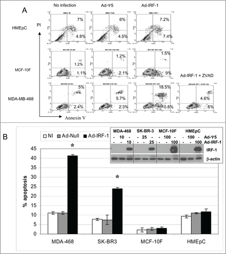
Figure 3. TNF-α and/or IFN-γ induces IRF-1 and enhances human breast cancer cell apoptosis. (A) The MDA-MB-468 and SK-BR-3 breast cancer cells were either cultured in medium alone or cultured with IFN-γ, TNF-α, or IFN-γ and TNF-α as described in Materials and Methods. Apoptosis was evaluated as described in Materials and Methods. (B) The MDA-MB-468 or SK-BR-3 breast cancer cells were either not infected or infected with the control Ad-Ψ5 or Ad-IRF-1. Infected cells were subsequently cultured with TNF-α and apoptosis was measured by FITC-conjugated annexin V and propidium iodide as described in Materials and Methods. Percent apoptosis indicated in the bar graphs represents the mean percent of gated cells in the early and late apoptotic quadrants. Standard deviation is indicated by error bars. * = p < 0.05 by unpaired t test. p < 0.01 for the MDA-MB-468/Ad-IRF-1 + TNF-α cohort compared with the MDA-MB-468/Ad-IRF-1 cohort and p < 0.0001 for the SK-BR-3/Ad-IRF-1 + TNF-α compared with SK-BR-3/Ad-IRF-1 breast cancer cohort. (C) The MDA-MB-468 and SK-BR-3 human breast cancer cells were either cultured in media, or cultured with TNF-α, IFN-γ, or the combination of TNF-α and IFN-γ as described in Materials and Methods. Cells were harvested after 24 h in culture and immunoblotting was performed as described in Materials and Methods.
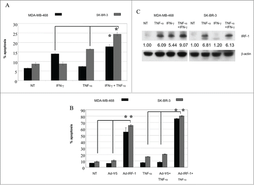
Figure 4. Abrogation of IRF-1 results in diminished IFN-γ and TNF-α induced apoptosis. (A) MDA-MB-468 breast cancer cells were either not transfected or transfected with a control siNeg, or siRNA to IRF-1. Untransfected cells were treated with the transfection reagent alone (T.R.) as an additional control. Cells were harvested and immunoblotting was performed as described in Materials and Methods. (B) Abrogation of IRF-1 results in diminished IFN-γ and TNF-α induced apoptosis. MDA-MB-468 human breast cancer cells were transfected with a control siNeg or siRNA to IRF-1. 24 h post transfection the cells were cultured with IFN-γ and TNF-α and cell death was evaluated as described in Materials and Methods. Standard deviation is indicated by error bars. *= p < 0.05 by unpaired t test.
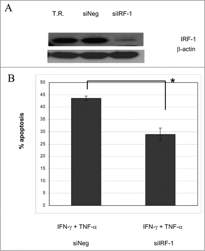
Figure 5. Ectopic expression of IRF-1 reduces NF-κB expression and activity in human breast cancer cells. (A) The nonmalignant human mammary epithelial cells HMEpC, the spontaneously immortalized nonmalignant human breast cell line MCF-10F, and the human breast cancer cell lines MDA-MB-468 and the SK-BR-3 were either uninfected or infected with the control Ad-Ψ5 or Ad-IRF-1 as previously described. Cellular lysates were prepared and immunoblotting for NF-κB p65 was performed as described in Materials and Methods. (B) The MDA-MB-468 and SK-BR-3 human breast cancer cells were co-transfected with the NF-κB responsive luciferase reporter construct pELAM-luc and β-gal plasmid pIEPlacZ. 24 h post transfection cells were either not infected or infected with the control Ad-Ψ5, Ad-IRF-1, or Ad-IκB super-repressor in duplicate. 24 h post infection luciferase activity was measured as described in Materials and Methods. (C) Uninfected cells or cells infected with the Ad-Ψ5 control or Ad-IRF-1 at the indicated MOIs were harvested 24 h post infection. Cellular lysates were used for immunoblotting as described in Materials and Methods.
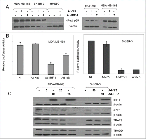
Figure 6. p65 protein expression is decreased in the nuclear fraction of the human breast cancer cell line MDA-MB-468. (A) The human breast cancer cells, MDA-MB-468, were either not treated or cultured with TNF-α, IFN-γ, or the combination of TNF-α and IFN-γ. 24 h post treatment, cells were harvested and nuclear and cytosolic fractions were isolated. (B) The spontaneously immortalized nonmalignant human breast cell line, MCF-10F, was either not treated or cultured with TNF-α, IFN-γ, or the combination of TNF-α and IFN-γ as described in Materials and Methods. 24 h post culture, cells were harvested and immunoblotting was performed as described in Materials and Methods. (C) The MCF-10F cells were either not treated or cultured with TNF-α, IFN-γ, or the combination of TNF-α and IFN-γ. 24 h post treatment, cells were harvested and nuclear and cytosolic fractions were isolated. Immunoblotting for NFκB p65 in nuclear and cytosolic fractions was performed as described in Materials and Methods.
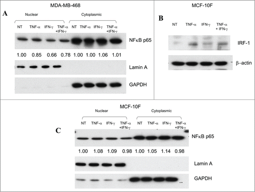
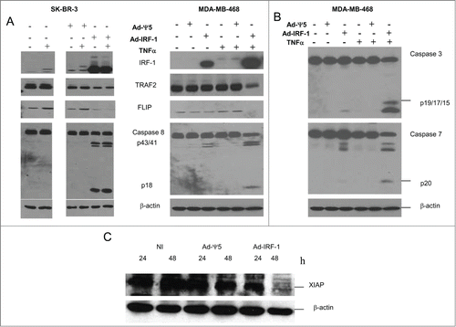
Figure 7. FLIP protein expression is suppressed by Ad-IRF-1 infection in human breast cancer cells. (A) SK-BR-3 and MDA-MB-468 human breast cancer cells were either uninfected or infected with Ad-Ψ5 or Ad-IRF-1 at a MOI of 50 and 25 respectively and cultured with or without 500 U/mL of TNF-α. After 24 h, cells were harvested and cellular lysates were loaded and utilized for immunoblotting as described in Materials and Methods. Multiple experiments were run on the same gel and at the same time, and the relevant results are presented. (B) MDA-MB-468 human breast cancer cells were either uninfected or infected with Ad-Ψ5 or Ad-IRF-1 at a MOI of 25 and cultured with or without 500 U/mL of TNF-α. After 24 h, cells were harvested and cellular lysates were loaded and utilized for immunoblotting as described in Materials and Methods. (C) XIAP expression is decreased in Ad-IRF-1 infected human breast cancer cells. MDA-MB-468 human breast cancer cells were either uninfected or infected with Ad-Ψ5 or Ad-IRF-1 and immunoblotting was conducted as described in Materials and Methods.