Figures & data
Figure 1. Metformin inhibits the proliferation and colony formation of prostate cancer cells. LNCaP and PC3 cells were treated with metformin (Met) at various concentrations (0, 0.625, 2.5, and 10 mM). (A) Cell proliferation was determined at 24, 48, or 72 h by the CCK-8 kit. (B-C) Colony formation was quantified 14 d after treatment with metformin by counting the colonies formed. *p < 0.05; **p < 0.01; §p < 0.001 vs the control.
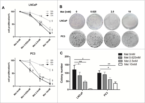
Figure 2. Metformin induces apoptosis of prostate cancer cells. LNCaP and PC3 cells were treated with metformin (Met) at various concentrations (0, 0.625, 2.5, and 10 mM). After 24 h, apoptosis was quantified by Annexin V-APC assay and PI staining. (A) Early apoptotic cells (lower right, LR) and late apoptotic cells (upper right, UR) are shown. (B) Histograms represent quantification of the rate of apoptosis. *p < 0.05; **p < 0.01; §p < 0.001 vs the control.
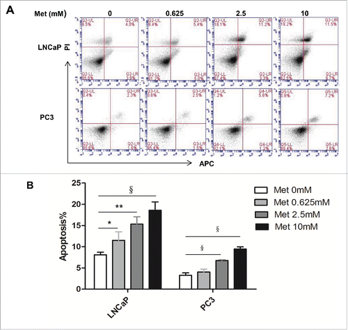
Figure 3. Metformin inhibits prostate cancer cell migration and invasion. (A-B) Cell migration was determined by a Boyden chamber assay (LNCaP) or a wound closure assay (PC3) following treatment with metformin (Met) for 48 h at the indicated concentrations (0, 0.625, 2.5, and 10 mM). The images are representative counted fields for LNCaP and wound closures for PC3. Data (mean ± SD) are expressed graphically as percentage of cells migrating. (C-D) The invasion assay was performed in Boyden chambers after 48 h in the presence of various concentrations of metformin (0, 0.625, 2.5, and 10 mM). Cells that invaded into the lower chamber were counted, and the images are representative counted fields. Data (mean ± SD) are expressed graphically as percentage of cells invading. *p < 0.05,**p < 0.01, §p < 0.001 vs the control.
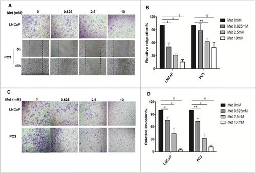
Figure 4. Metformin inhibits xenograft tumor growth in nude mice. Xenograft tumors were generated by subcutaneous implantation of PC3 cells (5 mice/group). Metformin treatment (125 mg/kg [Met 125] or 250 mg/kg [Met 250] daily) was initiated 15 d later, and the treatment continued 20 d. (A) PC3 xenograft tumors were measured every 3 d during treatment; mean tumor volumes over time are shown. (B-C) Photographs show the xenograft tumor-bearing mice and tumors by treatment group. (D) Average weights of excised tumors by treatment group are shown. *p < 0.05; **p < 0.01; §p < 0.001 vs the control. #p < 0.001, Met 125 vs Met 250.
![Figure 4. Metformin inhibits xenograft tumor growth in nude mice. Xenograft tumors were generated by subcutaneous implantation of PC3 cells (5 mice/group). Metformin treatment (125 mg/kg [Met 125] or 250 mg/kg [Met 250] daily) was initiated 15 d later, and the treatment continued 20 d. (A) PC3 xenograft tumors were measured every 3 d during treatment; mean tumor volumes over time are shown. (B-C) Photographs show the xenograft tumor-bearing mice and tumors by treatment group. (D) Average weights of excised tumors by treatment group are shown. *p < 0.05; **p < 0.01; §p < 0.001 vs the control. #p < 0.001, Met 125 vs Met 250.](/cms/asset/51d5c49f-d7bd-40fc-b506-af27b58325fc/kcbt_a_1156273_f0004_oc.gif)
Figure 5. Metformin increases PEDF expression in prostate cancer cells. LNCaP and PC3 cells were treated with metformin (Met) at different concentrations (0, 0.625, 2.5, and 10 mM) for 24 h. (A) Total RNA was isolated and reverse-transcribed for qPCR to estimate the levels of PEDF mRNA in LNCaP and PC3 cells. (B-C) The whole-cell proteins were analyzed by protein gel blot. Data represent mean ± SD. *p < 0.05; **p < 0.01; §p < 0.001 vs the control.
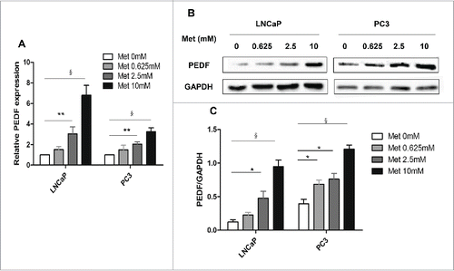
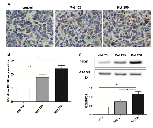
Figure 6. Metformin increases PEDF expression in vivo. (A) Paraffin sections of tumors excised from mice treated with vehicle (control), metformin 125 mg/kg (Met 125), or metformin 250 mg/kg (Met 250) were assessed for PEDF expression by immunohistochemical staining. PEDF-positive cells are highlighted with arrows (400× magnification). (B-D) PEDF levels were quantified by qPCR (B) and western blot (C&D). *p < 0.05; **p < 0.01; §p < 0.001 vs the control.