Figures & data
Figure 1. Characteristics and differentiation potential of MSCs derived from umbilicalcord tissues. (A) Morphology of UC-MSCs Magnification:×100. After chondrogenic differentiation conditions, MSCs differentiate into chondrogenic-like cells and immunohistochemically stained positive for type II Collagen; ×100. After osteogenic-specific induction, the MSCs were stained with Alizarin red; ×100. After inducing adipogenic differentiation, the cells showed many small lipid vacuoles, as assessed by Oil Red O staining;×100. (B) Flow cytometric analysis showing the MSC cells surface antigens: positive for mesenchymal lineage markers (CD44, CD73, CD90 and CD105), negative for haematopoietic and endothelial markers (CD31, CD34 and CD45), and negative for HLA-DR.
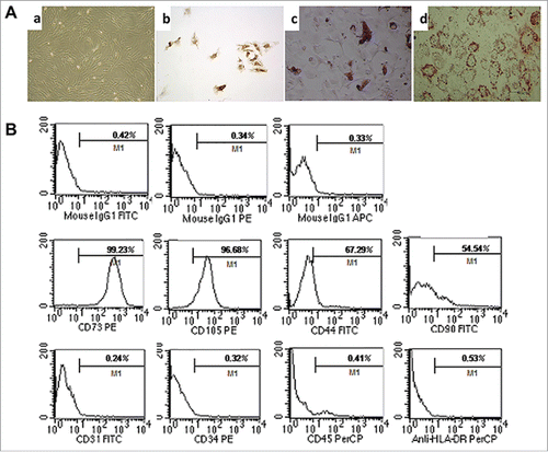
Figure 2. HNF4α stably expressed in MSCs. (A) The transduction efficiency of MSCs infected with lentiviral vectors was assessed based on the GFP expression in MSCs by immunofluorescence staining, and more than 90% of MSCs stably expressed GFP; (B) Real-time PCR showed that the HNF4α mRNA expression was significantly up-regulation in MSC-HNF4α compare with MSC (p < 0.01); (C)Western blotting indicated that the HNF4α protein expression was elevated.
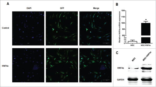
Figure 3. MSC-HNF4α inhibited HCC proliferation, migration and invasion. (A) Upper panel: CCK-8 assay showed that the OD value of SK-Hep-1 and HepG2 cells cultured with 50% MSC-HNF4α conditioned media was significantly decreased as compared to LO2 or MSC-conditioned media. Lower panel: Effect of conditioned-media on LO2 proliferation, no statistically significant difference was observed among 3 groups; (B) The colony formation assay showed that the proliferation of SK-Hep-1 and HepG2 cells treated with MSC-HNF4α conditioned media was significantly lower than that of the control group(L02) and MSC group; (C) The subcutaneous tumorigenicity assay showed that the weight and volume of SK-Hep-1 tumors treated with MSC-HNF4α were significantly decreased compared with those of the control group(NS) and MSC group; (D) The Matrigel invasion assay showed that MSC-HNF4α-conditioned medium significantly inhibits SK-Hep-1 and HepG2 cells invasion in vitro; (E) Immunofluorescence staining showed lower expression of MMP2 and MMP9 in HCC tissues (SK-Hep-1) following MSC-HNF4α treatment compared with the controls.×400; (F)Cell apoptosis assay showed that the different in each group was notstatistically significant. (*P < 0.05).
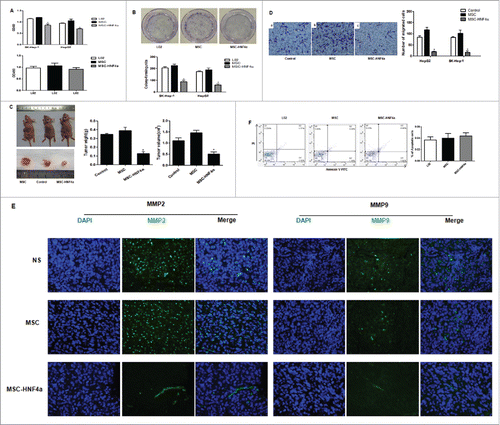
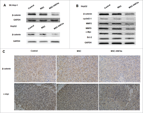
Figure 4. MSC-HNF4α inhibited HCC proliferation and invasion by inhibiting the Wnt/β-catenin signaling pathway. HCC cells were cultured with culture medium for 48 h, (A) protein gel blotting assay for β-catenin in SK-Hep-1 and HepG2 was downregulated when cells were treated with MSC-HNF4α conditioned media. (B) Target genes of the Wnt/β-catenin pathways, β-catenin, cyclin D1, MMP2, MMP9 and c-Myc were also down-regulated in MSC-HNF4α group but Blc-2 did not demonstrate any significant changes between each group. (C) Expression of β-catenin and c-Myc in tumor were clearly decreased in MSC-HNF4a group by immunohistochemical assay.