
Figures & data
Figure 1. Cerebrospinal fluid (CSF) cytological examination showed presence of abnormal plasma cells featuring enlarged and hyperchromatic nuclei, nuclear contour irregularity and occasional cells with binucleation. (Giemsa stain, original magnification, x 400).
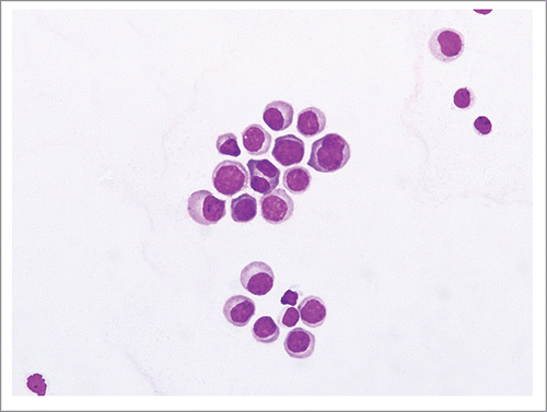
Figure 2. Flow cytometric analysis of the CSF specimen demonstrating plasma cells (PC). The PC are of intermediate side scatter and are CD45 positive (A), CD 38 brightly positive, CD 19 negative and CD 138 positive (B and C). They are also dimly positive for CD 28 and are CD 27 negative (D); this is a feature of aberrant PC. (Van Dongen et al Leukemia 2012).
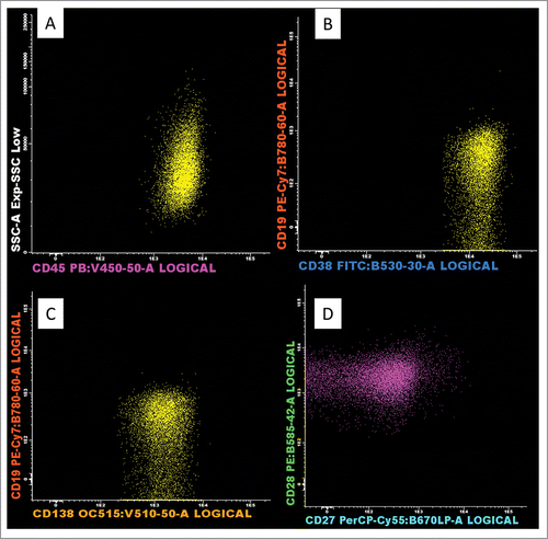
Table 1. Treatment and outcomes of CNS-MM in the era of NAs.