Figures & data
Figure 1. Histological and immunophenotype of parental (patient) tumor, its derived TSG and lung metastasis. (A-E) Hematoxylin and eosin (H&E) staining demonstrated similar histology among parental tumor, primary first generation (1st) TSG under the renal capsule and its derived lung metastasis, and primary subcutaneous (subQ) tumor from sixth generation TSG and its derived lung metastasis. IHC revealed similar expression of human-specific nuclear antigen Ku70 (F-J) and typical markers of pRCC including AMACR (K-O) and Pax-8 (P-T) among the parental tumor, primary TSGs and metastases. No expression of CD117 was detected (U-Y). Magnification of all images is 200×. Dotted lines in (H) (J) (M) (O) (R) (T) (W) and (Y) delineate metastatic tumor cells vs. mouse tissue in the lung.
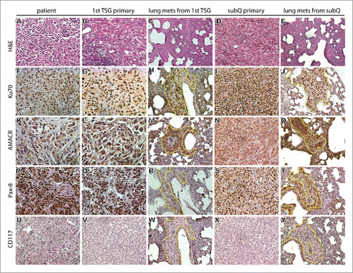
Table 1. Engraftment rates of TSGs and metastasis rate.
Figure 2. Detection of MET mutation by direct sequencing of tumor tissue DNA and plasma ctDNA. The same point mutation [T-3997C(M1268T)] in the MET kinase domain (A) was detected in patient tumor tissue (B), 1° (first generation) subrenal TSG (C) and ctDNA from mice carrying 4° (fourth generation) subrenal TSGs (D).
![Figure 2. Detection of MET mutation by direct sequencing of tumor tissue DNA and plasma ctDNA. The same point mutation [T-3997C(M1268T)] in the MET kinase domain (A) was detected in patient tumor tissue (B), 1° (first generation) subrenal TSG (C) and ctDNA from mice carrying 4° (fourth generation) subrenal TSGs (D).](/cms/asset/c443ce2b-b321-4c3e-82c8-0d2131e1cc8f/kcbt_a_1219816_f0002_c.gif)
Figure 3. Regression of primary tumor induced by cabozantinib in TSG-RCC-030. Subcutaneous tumor growth was measured in control and cabozantinib-treated mice. Tumor volume significantly decreased over the time course of 21 d in treated mice while continuously increasing in control mice (A). The specific tumor growth rate was negative in treated mice and positive in control mice (B). IHC demonstrated strong expression of the proliferation marker Ki67 in a subset of cells (arrows) and phosphorylated MET as well as its target, phosphorylated ERK, in majority of tumor cells in control but not in treated mice (C). Data points represent mean+/− SD. *p < 0.05 by Student's t-test. Magnification of all images is 200×.
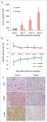
Figure 4. Inhibition of TSG-RCC-030 lung metastasis by cabozantinib. The level of human GAPDH in the lung tissue of control mice was significantly higher than in cabozantinib-treated mice as assessed by qPCR using human-specific GAPDH primers (A). Tumor cells expressing human-specific nuclear antigen Ku70 were detected only in control but not treated mice (B). These metastatic tumor cells also expressed Pax-8, a typical marker of pRCC (B). Data points represent mean+/− SD. *p < 0.05 by Student's t-test. Magnification of images is 200×.
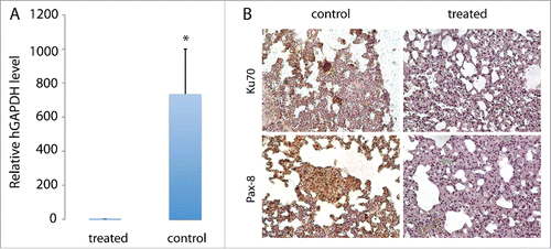
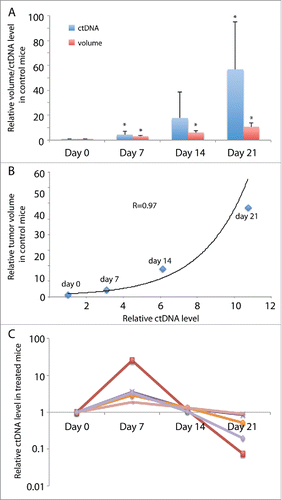
Figure 5. ctDNA levels in mice bearing TSG-RCC-030 correlated with tumor volume and responded to cabozantinib treatment. ctDNA levels were measured by qPCR using allele-specific primers that amplified the mutant MET. ctDNA levels increased continuously over time, reaching a 57-fold higher level at the end of the experiment in control mice (A). In control mice, tumor volume correlated with an exponential increase in ctDNA level with a correlation coefficient of 0.97 by Pearson Correlation analysis (B). In treated mice, ctDNA levels increased at 7 d after initiation of treatment and thereafter decreased to different extents in individual mice (C). Data points represent mean+/− SD in (A), mean in (B) and individual in (C). *p < 0.05 by Student's t-test.