Figures & data
Figure 1. Clinically relevant inhibitors of the isoprenoid biosynthetic pathway (IBP) (a) Statins inhibit HMG CoA reductase, consequently reducing the entire IBP. Nitrogenous bisphosphonates inhibit FPP synthase (FDPS), consequently reducing FPP, GGPP, farnesylation, and geranylgeranylation. Our novel isoprenoid bisphosphonate compounds (b) DGBP and (c) GGOHBP inhibit GGPP synthase (GGDPS), consequently reducing only GGPP and protein geranylgeranylation.
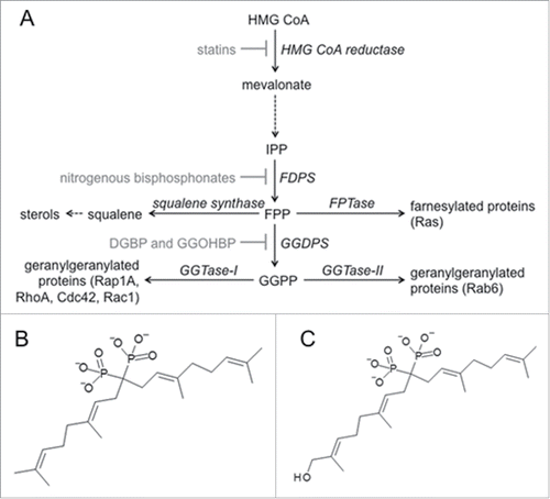
Figure 2. Novel compound GGOHBP inhibits Rap1A and Rab6 geranylgeranylation without altering Ras farnesylation (a) GGDPS enzyme inhibition and (b) MTT activity of the luciferase-expressing PC-3 cell line following 48 hr treatment with the indicated concentrations of each compound. Representative Western blot analysis of (c) Ras, (d) Rap1A, and (e) Rab6 following 48 hr treatment with the indicated concentrations of each compound in the luciferase-expressing PC-3 cell line. The anti-Ras and anti-Rab6 antibodies detect the unprenylated (aqueous) and prenylated (detergent) forms of Ras and Rab6, requiring separation by TX-114 detergent. The anti-Rap1A antibody detects the unprenylated form of Rap1A. Experiments were performed in triplicate. Error bars indicate standard deviation.
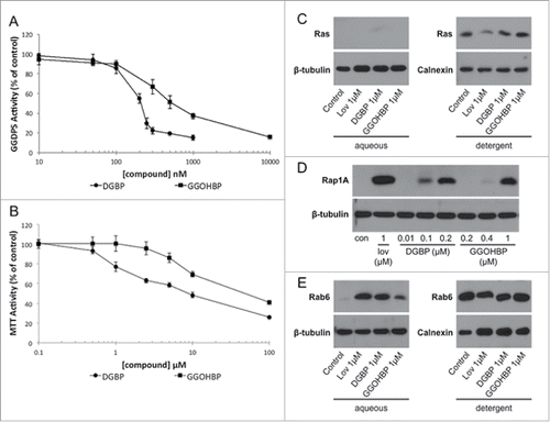
Table 1. Distribution of tumors as determined by ex vivo BLI. Sites of metastasis are ordered from most common to least common presentation in all mice.
Figure 3. Mice receiving daily treatment of GGOHBP have a reduced whole body tumor burden as shown by BLI (a) Representative weekly BLI of control mouse C4M5 and treated mouse C2M2. The red dashed line indicates the missing photographs from weeks 5 and 6 due to a required temporary switch to the AMI instrument. Control mouse C4M5 died during imaging on day 75 (week 11) whereas treated mouse C2M2 was removed from the study on day 117 (between weeks 16 and 17). (b) Whole body photon counts averaged within the vehicle-control (solid line) and GGOHBP (dashed line) treated groups during weeks 1-7 when all mice were alive. (c) Whole body photon counts for the entirety of the study (weeks 1-19) averaged within the control and treated groups. Statistical significance indicated as * (P < 0.05) as determined by Student's T Test. Error bars indicate standard deviation. N = 9 control vehicle-treated mice. N = 8 GGOHBP treated mice.
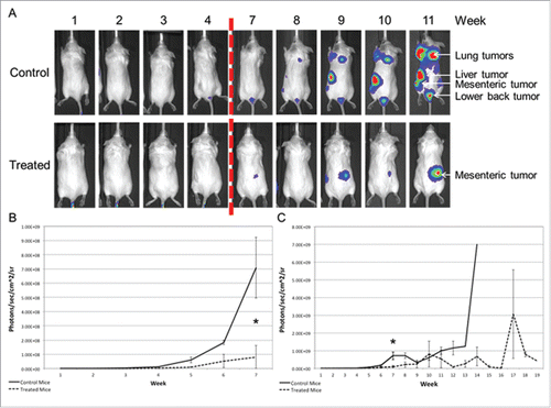
Figure 4. GGOHBP significantly slowed tumor development and prolonged overall survival Time until a whole body tumor burden of greater than (a) 107, (b) 108, or (c) 109 photon counts was detected for each mouse. (d) Weight of mice averaged within the control and treated groups. (e) Kaplan-Meier survival curve. Statistical significance indicated as * (P < 0.05) and ** (P < 0.005) as determined by Student's T Test. Error bars indicate standard deviation.
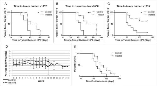
Figure 5. Rap1A geranylgeranylation is not altered in non-tumor burdened heart, liver, kidney, adrenal glands, and lung tissues of GGOHBP treated mice Representative Western blot analysis of Rap1A in non-tumorous (a) heart, (b) liver, (c) kidney, (d) adrenal glands and (e) lung tissues from control and treated mice. Control mouse C4M5 had too high of a lung tumor burden to collect non-tumorous lung tissue and so was not included. The anti-Rap1A antibody detects the unprenylated form of Rap1A as shown by the positive control GGOHBP treatment in luciferase-expressing PC-3 cells.
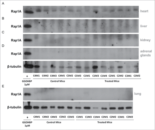
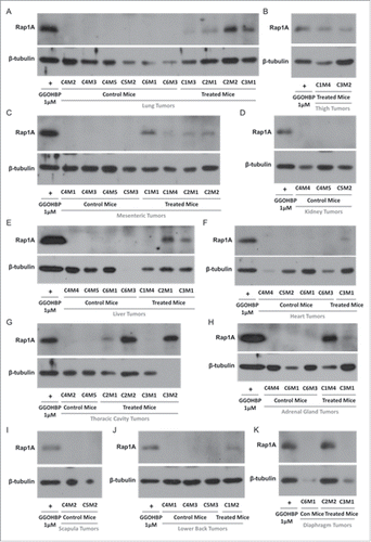
Figure 6. Rap1A geranylgeranylation is reduced in all soft tissue tumors of GGOHBP treated mice Representative Western blot analysis of Rap1A in (a) lung tumors, (b) thigh tumors, (c) mesenteric tumors, (d) kidney tumors, (e) liver tumors, (f) heart tumors, (g) thoracic cavity tumors, (h) adrenal gland tumors, (i) scapula tumors, (j) lower back tumors, and (k) diaphragm tumors of control and treated mice. The anti-Rap1A antibody detects the unprenylated form of Rap1A as shown by the positive control GGOHBP treatment in luciferase-expressing PC-3 cells.