Figures & data
Figure 1. Schematic of the study design. This study was designed to test the central hypothesis that the protective effects of Se against CICC are APN independent. Flowchart illustrates the time-points at which DSS and DMH were administered in the DSS+DMH group, the time period during which se-diet was administered and the time-point at which the animals were killed and analyzed for CICC (see results).
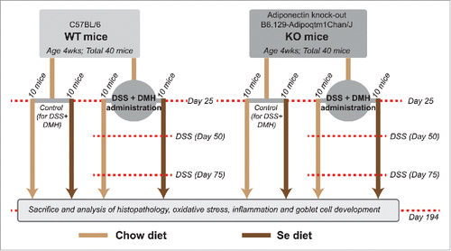
Figure 2. Association of Se mediated reduction of CICC with Se and Selenoprotein accumulation and reduction of oxidative stress in colon tissues. (a) Reduction in tumor numbers and tumor area. (i) Representative methylene blue stained tissues. (ii) Comparison of colon tumor numbers in KO and WT mice on Se and chow diets (N = 10 per group). (iii) Comparison of colon tumor area (mm2) in KO and WT mice on Se and chow diets (N = 10 per group). Significance in difference was calculated using one way ANOVA. *p < 0.05 between WT and WT+Se, #p < 0.01 between KO and KO+Se, €p < 0.05 between WT+Se and KO+Se, ¥p < 0.05 between WT mice on chow diet and KO mice on chow diet. (b) Increased Se content in colon tissues of mice on Se diet. Mass spectrometry data showing Se content of the colon tissues in mice on control and Se diet. *p < 0.05, significance in difference in Se content in colon tissues of WT or KO mice (either control group or DSS+DMH administered group) on chow diet vs. Se diet was calculated using one-way ANOVA, N = 10 per group (c) Se-mediated reduction in oxidative stress in colon tissues. Bars showing a quantitative comparison between 4HNE and Nitrotyrosine levels. *p < 0.05, significance in difference between mice on control vs. Se diet was determined using one way ANOVA, N = 10 per group (d) Effect of Se diet on selenoproteins activity and Gpx1 and Gpx2 protein levels: i. Bar graph representing the Gpx activity (nmol/min/mg protein) in the colon tissue sections in control group mice and DSS+DMH administered mice. *p < 0.05 between mice on Se and regular chow diet within same treatment group and genotype, N = 10 per group ii. Bar graphs derived from Western Blot data (SI- 2) showing a quantitative comparison of Gpx-1 and Gpx-2 protein levels in control group mice and DSS+DMH administered mice, N = 10 per group. One way ANOVA was used to determine the significance in difference between mice on Se and regular chow diet within the same treatment group with same genotype.
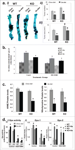
Figure 3. Effect of Se diet on inflammation, apoptosis and goblet cell differentiation. (A) Effect on inflammation i) Histopathology Staining and Scores. Left panel. Representative hematoxylin and eosin stained colon tissue sections of control and DSS+DMH treated mice. Right panel. Histopathology scores representing a culmination of inflammation, immune cell infiltration and degree of tumor, derived from HE stained images. One way ANOVA was used to determine significance in difference (N = 10 per group). *p < 0.05 between mice on Se and regular chow diets with in the same genotype and treatment group, #p < 0.03 between WT mice and KO mice on regular chow diet, **p < 0.04 between WT mice on regular chow diet and Se diet, ***p < 0.01 KO mice on control and Se diet. ii) Effect on markers of inflammation. Bar graphs representing the levels of secreted cytokines from the colon tissue section. *p < 0.05, #p < 0.01, One way ANOVA was used to determine significance in the difference between mice of a particular genotype and treatment on Se and regular chow diet (N = 10 per group). (B) Effect of Se diet on apoptosis. Upper panel. Images of representative colon tissues showing the TUNEL positive epithelial cells (brown color) in the control group and the non-tumor and tumor regions of colon tissue sections of DSS+DMH administered KO and WT mice on control or Se diet. The tissues were counterstained with methyl green. Lower panel, Graph generated from the TUNEL stained images representing ratio of the number of TUNEL positive cells to the total epithelial cells (degree of apoptosis). One way ANOVA was used to determine significance in difference; N = 10 per group. #p < 0.04 and **p < 0.05 between mice same genotype and treatment group on Se and regular chow diet, *p < 0.05 between WT+Se and KO+Se, (C) Effect of Se diet on goblet cell counts. Left panel, Representative Alcian Blue and Nuclear Fast Red stained colon tissue images. Right panel. Quantification of goblet and epithelial cells in colon tissues in mice on control or Se diet. One way ANOVA (N = 10 per group) was used to determine significance in difference, *p < 0.05 between mice same genotype and treatment group on Se and regular chow diet. (D) Quantitative comparison of Math-1 and Muc-2 protein levels generated from Western Blots (representative images shown in SI- 4). *p < 0.05, significance of difference between mice on regular chow diet and Se diet was determined using one way ANOVA.
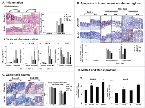
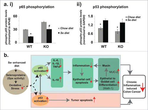
Figure 4. Selenium's effects on p65 and p53 phosphorylation and the protection model against CICC. (a) i) Comparisons in p65 phosphorylation in DSS+DMH administered mice on regular chow diet and Se diet. Ratios of phospho p65 to total p65 levels were derived from the Western Blot data (representative image shown in SI-5). ii) Comparisons in p53 phosphorylation in DSS+DMH administered mice on regular chow diet and Se diet. Ratios of phospho p53 to GAPDH levels were derived from the Western Blot data (representative image shown in SI-6). *p < 0.05 and #p < 0.01, significance in difference between mice on control and Se diets were determined using one way ANOVA, (b) Selenium protection model: Se-mediated reduction of inflammation and increase in tumor apoptosis in colon tissues of DSS+DMH administered mice. According to this model, one mode of Se's protection involves the downregulation of inflammation that is driven by decreased phosphorylation of p65 global transcription factor and which results in decrease of pro-inflammatory cytokines IL-6, TNFα and IL-1β and increase in anti-inflammatory cytokine IL-10. The decrease in inflammation reduces epithelial cell apoptosis in non-tumor regions and thereby supports the epithelial to goblet cell differentiation (mediated by increase in Math-1) resulting in higher production of mucin (mediated by increase in Muc-2), a glycoprotein that in turn has protective effects against colon cancer. In the second mode, Se simultaneously results in increased tumor apoptosis possibly by Bax pathway (suggested by increase in cleaved caspase 9 levels), which in turn mediated by increase in phosphorylation of the tumor suppressor p53. Gray solid arrows represent mechanisms that have been established previously (and cited in the text). Red arrows (upward, increase and downward, decrease) are results reported in the current study. Our results suggest that Se mediated drop in oxidative stress regulates the cross-talk between p65 and p53; this molecular mechanism underlying this regulation remains unknown (dashed arrows) and will be elucidated in our future studies.