Figures & data
Figure 1. Numb expression in ESCC tissues and cell lines. (A) Immunohistochemistry (IHC) on tissue microarrays was used to measure Numb expression and upregulation in ESCC tissues. classes 1-3 correspond directly to Numb staining intensity. Images on left and right are magnified 100 × and 400 ×, respectively. (B) Upregulation of Numb protein in ESCC tissue compared with corresponding non-tumor tissue was confirmed by western blot. A representative image is shown. (C) Numb protein expression was evaluated by western blot in the different ESCC cell lines of KYSE30, KYSE140, EC9706, EC109, EC108 and TE-1, and normal esophageal tissue.
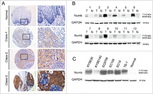
Table 1. Association between Numb protein expression and clinical characteristics in ESCC samples.
Figure 2. Increased Numb expression in ESCC tissue correlates with poor prognosis in patients. Numb protein levels in ESCC and adjacent normal tissues from 193 patients with complete long-term follow-up were measured by IHC. Kaplan-Meier analysis revealed that high Numb expression correlated with a significantly shorter (A) EFS (P = 0.015, log-rank test) and (B) OS time (P = 0.016, log-rank test) for all patients. p < 0.05 was considered significant.
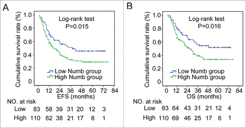
Table 2. Univariate analysis of influence of clinicopathological characteristics on ESCC patient prognosis.
Table 3. Cox multivariate analysis of influence of clinicopathological characteristics on ESCC patient prognosis.
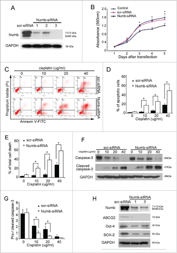
Figure 3. siRNA knockdown of endogenous Numb in KYSE30 cells inhibits proliferation, promotes apoptosis, and downregulates cancer stem cell biomarker expression. (A) Downregulation of Numb expression in KYSE30 cells using Numb-siRNA. (B) Using the MTT assay, it was determined that downregulation of Numb suppresses KYSE30 cell growth. (C-E) Following transfection with Numb-siRNA, KYSE30 cells were treated with a range of concentrations of cisplatin for 24 hours. Flow cytometry was performed to quantify the number of apoptotic cells and total dead cells following treatment. KYSE30 cells with lower expression of Numb had significantly more apoptotic cells and total dead cells than the negative control. Data are shown as mean ± SD and are from 3 independent experiments. (F-G) Western blots were performed to assess caspase-3 cleavage in KYSE30 cells following treatment with cisplatin. Decreased expression of Numb was associated with increased cleavage of caspase-3. (H) Decreased Numb expression resulted in lower levels of expression of Oct-4, Sox-2 and Nanog, which are biomarkers of cancer stem cells. All data are from at least 3 independent experiments. Differences were considered statistically significant at p < 0.05 versus the control.