Figures & data
Figure 1. Combination treatment with L-arginine and α-PD-L1 antibody significantly increased survival in mice bearing in situ and metastatic osteosarcoma. (A) The survival curves of mouse bearing in situ osteosarcoma were monitored. (B) The survival curves of mice with metastatic osteosarcoma. n = 10/group. **P < 0.01, ***P < 0.001, and **** P < 0.0001.
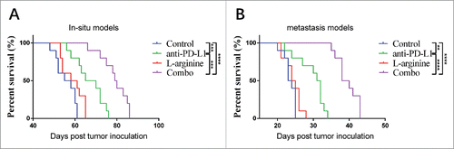
Figure 2. Combination treatment with L-arginine and α-PD-L1 antibody increased the proportion of CD8+ T-cells and CD86+ CD11c+ dendritic cells in the spleen of mice bearing in situ osteosarcoma. (A) Representative data of proportions of CD8+ T-cells. (B) Pooled data of proportions of CD8+ T-cells in the spleen in different groups. n = 5/group. (C) Representative data of proportions of mature dendritic cells. (D) Pooled data of proportions of mature dendritic cells in the spleen in different groups. n = 5/group. *** P < 0.001, and **** P < 0.0001. Data were presented as mean ± SEM.
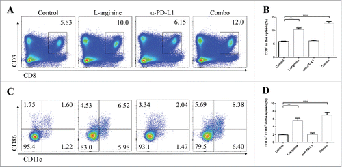
Figure 3. Combined treatment significantly elevated the number and activity of CD8+ T-cells in tumor-draining lymph nodes. (A) Representative data of proportions of CD8+ T-cells in TDLNs. (B) Pooled data of proportions of CD8+ T-cells in TDLNs in different groups. Representative data of proportions of IFN-γ+ (C), granzyme-B+ (E), and perforin+ (G) cells in CD8+ cells from TDLNs. Pooled data of frequencies of IFN-γ+ (D), granzyme-B+ (F), and perforin+ (H) cells in CD8+ cells in TDLNs from different groups. n = 5/group. * P < 0.05, ** P < 0.01, *** P < 0.001, and **** P < 0.0001. Data were presented as mean ± SEM.
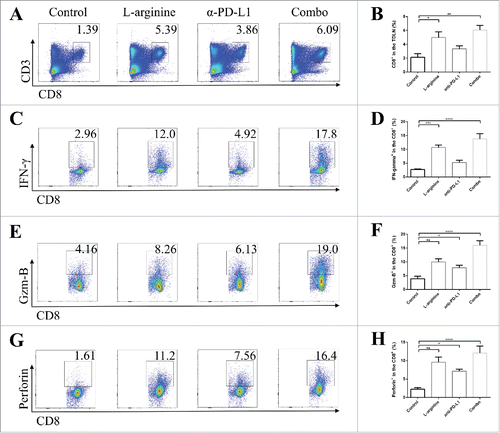
Figure 4. Combined treatment significantly elevated the number and activity of CD8+ T-cells in orthotopic tumors. (A) Representative data of proportions of CD8+ T-cells in tumor. (B) Pooled data of proportions of CD8+ T-cells in tumor in different groups (calculated from CD8+/total events). Representative data of proportions of PD-1+ (C) and IFN-γ+ (E) cells in intratumoral CD8+ cells. Pooled data of frequencies of PD-1+ (D) and IFN-γ+ (F) cells in intratumoral CD8+ cells from different groups. n = 5/group. * P < 0.05, ** P < 0.01, *** P < 0.001, and **** P < 0.0001. Data were presented as mean ± SEM.
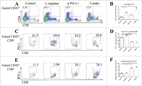
Figure 5. Combined treatment promoted immune response in pulmonary metastasized osteosarcoma. (A) Representative data of frequencies of CD8+ T-cells in metastasized osteosarcoma. (B) Pooled data of proportions of CD8+ T-cells in tumor in different groups (calculated from CD8+/total events). Representative data of proportions of PD-1+ (C) and IFN-γ+ (E) cells in pulmonary intratumoral CD8+ cells. Pooled data of frequencies of PD-1+ (D) and IFN-γ+ (F) cells in intratumoral CD8+ cells from different groups. n = 5/group. * P < 0.05, ** P < 0.01, *** P < 0.001, and **** P < 0.0001. Data were presented as mean ± SEM.
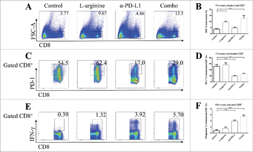
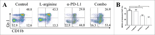
Figure 6. L-arginine plus α-PD-L1 antibody inhibited MDSC proliferation in orthotopic osteosarcoma models. (A) Representative data of frequencies of CD11b+ Gr-1+ MDSCs in intratumoral CD45+ cells. (B) Pooled data of proportions of MDSCs in tumor in different groups (calculated from CD11b+ Gr-1+/CD45+). n = 5/group. * P < 0.05 and ** P < 0.01. Data were presented as mean ± SEM.