1,147
Views
13
CrossRef citations to date
0
Altmetric
Research Paper

Direct measurement of hypoxia in a xenograft multiple myeloma model by optical-resolution photoacoustic microscopy

, , , , , & show all
Pages 101-105 | Received 09 Nov 2016, Accepted 18 Dec 2016, Published online: 26 Jan 2017

Figures & data

Figure 1. Optical-resolution photoacoustic microscopy (OR-PAM) for in vivo imaging of oxygenation and vascularization in the bone marrow. (A) Schematic diagram of the OR-PAM imaging system. AL, acoustic lens; CL, correction lens; DAQ, data acquisition; DM, dichroic mirror; FC, fiber coupler; M, mirror; ND, neutral density filter; PD, photodiode; PH, pinhole; PR, prism; SMF, single-mode fiber; SO, silicone oil; UT, ultrasonic transducer. (B) Imaging regions in the mouse skull.
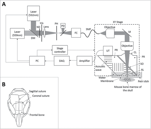
Figure 1. Optical-resolution photoacoustic microscopy (OR-PAM) for in vivo imaging of oxygenation and vascularization in the bone marrow. (A) Schematic diagram of the OR-PAM imaging system. AL, acoustic lens; CL, correction lens; DAQ, data acquisition; DM, dichroic mirror; FC, fiber coupler; M, mirror; ND, neutral density filter; PD, photodiode; PH, pinhole; PR, prism; SMF, single-mode fiber; SO, silicone oil; UT, ultrasonic transducer. (B) Imaging regions in the mouse skull.

Figure 2. The sO2 level and blood vessel density are inversely correlated with the tumor burden. (A) Images of sO2 levels acquired by OR-PAM and the corresponding fluorescence microscopy images. The mouse brain was imaged at Day 1, Day 14, and Day 28 after intravenous injection of MM cells. Scale bars: 1 mm. (B) Designated regions of interest (ROI) of mouse 1 and mouse 2 used for analysis denoted as normal areas, regions A and B, and tumor areas, regions C and D. (C) Tumor progression evaluated as the tumor area (mm2) measured at Days 1, 14, and 28 post MM cell injection (mean ± SD), n = 4. (Di) Comparison of average relative total sO2 level between tumor and normal regions over the course of 4 weeks and normalized to Day 1 post MM injection (mean ± SD), n = 4. (Dii) Correlation between the oxygenation (total sO2) and the tumor area (mm2) with correlation coefficient R2 = 0.8096. (Ei) Comparison of average blood vessel density between tumor and normal regions over the course of 4 weeks and normalized to Day 1 post MM injection (mean ± SD), n = 4. (Eii) Correlation between the blood vessel density and the tumor area (mm2) shows a correlation coefficient R2 = 0.7757.
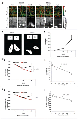
Figure 2. The sO2 level and blood vessel density are inversely correlated with the tumor burden. (A) Images of sO2 levels acquired by OR-PAM and the corresponding fluorescence microscopy images. The mouse brain was imaged at Day 1, Day 14, and Day 28 after intravenous injection of MM cells. Scale bars: 1 mm. (B) Designated regions of interest (ROI) of mouse 1 and mouse 2 used for analysis denoted as normal areas, regions A and B, and tumor areas, regions C and D. (C) Tumor progression evaluated as the tumor area (mm2) measured at Days 1, 14, and 28 post MM cell injection (mean ± SD), n = 4. (Di) Comparison of average relative total sO2 level between tumor and normal regions over the course of 4 weeks and normalized to Day 1 post MM injection (mean ± SD), n = 4. (Dii) Correlation between the oxygenation (total sO2) and the tumor area (mm2) with correlation coefficient R2 = 0.8096. (Ei) Comparison of average blood vessel density between tumor and normal regions over the course of 4 weeks and normalized to Day 1 post MM injection (mean ± SD), n = 4. (Eii) Correlation between the blood vessel density and the tumor area (mm2) shows a correlation coefficient R2 = 0.7757.

Reprints and Corporate Permissions

Please note: Selecting permissions does not provide access to the full text of the article, please see our help page How do I view content?

To request a reprint or corporate permissions for this article, please click on the relevant link below:

Academic Permissions

Please note: Selecting permissions does not provide access to the full text of the article, please see our help page How do I view content?

Obtain permissions instantly via Rightslink by clicking on the button below:

If you are unable to obtain permissions via Rightslink, please complete and submit this Permissions form. For more information, please visit our Permissions help page.