Figures & data
Figure 1. Antitumor effect of combinatorial treatment of H101 and DTIC in SP6.5 xenografted mice. (A) Average tumor volume of the mice bearing SP6.5 UM xenograft after treatment with PBS, H101, DTIC, and H101+DTIC. Values represent the means ± SD. **P<0.01 compared with H101+DTIC group. (B) Kaplan–Meier survival curves for overall survival of the tumor-bearing mice.*P<0.05compared to H101+DTIC group.
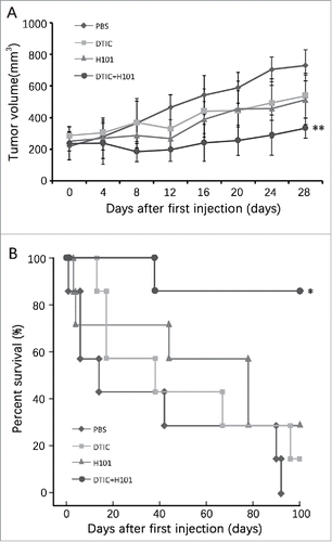
Figure 2. Down-regulation of MGMT expression by H101 in UM cells. (A) Western blot analysis of MGMT expression 48 hours after PBS, DTIC, H101, and DTIC+H101 treatment in SP6.5 and VUP cells. (B) Western blot analysis of MGMT expression 48 hours after PBS, DTIC, H101, and DTIC+H101 treatment in human normal pigment epithelial cells RPE.
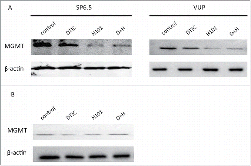
Figure 3. p53 and not p300 is involved in H101-induced MGMT downregulation. (A) Western blot analysis of E1A and p300 expression 48 h after PBS, DTIC, H101, and DTIC+H101 treatment in SP6.5 and VUP cells. (B) Western blot analysis of p53 expression 48 h after treatment in SP6.5 and VUP cells.
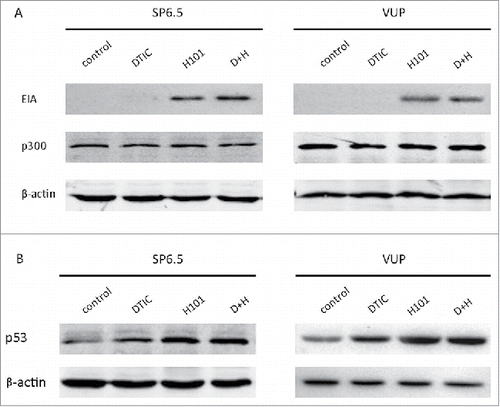
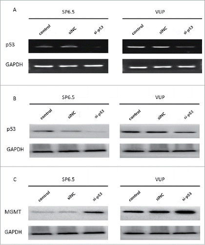
Figure 4. siRNA transfection downregulated p53 expression and resulted in an upregulation of MGMT. (A) PCR analysis of p53 expression in VUP and SP6.5 cells 48 h after transfection with p53-specific siRNA (si-p53), negative control siRNA (siNC) or PBS control (control). (B) Western blot analysis of p53 expression in VUP and SP6.5 cells 48 h after transfection. (C) Western blot analysis of MGMT expression in VUP and SP6.5 cells 48 h after transfection.