
Figures & data
Figure 1. Effects of PA after oral administration of 10 and 25 mg/kg doses for 15 d. Data represented as mean ± SD (n = 6). Statistically significant differences were observed between carcinogen control and test groups [one way-ANOVA followed by Bonferroni multiple comparison test (**p < 0.01, *p < 0.001)].
![Figure 1. Effects of PA after oral administration of 10 and 25 mg/kg doses for 15 d. Data represented as mean ± SD (n = 6). Statistically significant differences were observed between carcinogen control and test groups [one way-ANOVA followed by Bonferroni multiple comparison test (**p < 0.01, *p < 0.001)].](/cms/asset/e9e5836e-2a08-4f02-9b06-70196fc8dad4/kcbt_a_1310341_f0001_b.gif)
Table 1. Effects of PA on oxidative stress parameters in colon after 10 and 25 mg/kg doses for 15 d.
Figure 2. Effect of PA on enzyme levels in plasma after 10 and 25 mg/kg doses for 15 d. Data represented as mean ± SD (n = 6). Statistically significant differences were observed between carcinogen control and test groups [one way-ANOVA followed by Bonferroni multiple comparison test (*p < 0.001, **p < 0.01)].
![Figure 2. Effect of PA on enzyme levels in plasma after 10 and 25 mg/kg doses for 15 d. Data represented as mean ± SD (n = 6). Statistically significant differences were observed between carcinogen control and test groups [one way-ANOVA followed by Bonferroni multiple comparison test (*p < 0.001, **p < 0.01)].](/cms/asset/4cc9fd62-412e-481b-b472-21148eebceee/kcbt_a_1310341_f0002_b.gif)
Table 2. Effects of PA on (A) COX-2, IL-2 and IL-6 (B) caspase 3 in colon carcinogenic tissue after oral administration of 10 and 25 mg/kg for 15 d.
Figure 3. Gene expression levels of proinflammatory cytokines like (A) IL-6 and (B) COX-2 after PA administration in DMH treated rats. Data represented as mean ± SD (n = 6). Statistically significant differences were observed between carcinogen control and test groups [Paired T-test, (*p<0.001)].
![Figure 3. Gene expression levels of proinflammatory cytokines like (A) IL-6 and (B) COX-2 after PA administration in DMH treated rats. Data represented as mean ± SD (n = 6). Statistically significant differences were observed between carcinogen control and test groups [Paired T-test, (*p<0.001)].](/cms/asset/b3de94a4-7df8-47f0-bfeb-e581c10a16b6/kcbt_a_1310341_f0003_b.gif)
Figure 4. The colonic pathological changes in DMH-induced CC rats (Scale bar 50 µm). Tumoral vacuoles were prominent in DMH group which was absent after 5-FU and PA administration. (Cr- Crypts, NE- Normal epithelium, CL− Colon lumen, TA- Tubular adenoma, TD- Tumoral deposits, Ly- Lymphatics (lined by endothelium), TS- Tumor stroma).
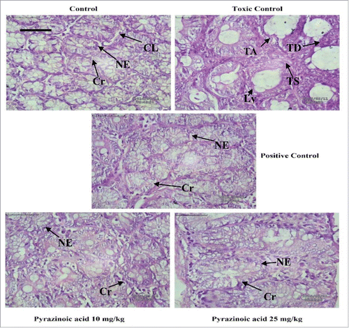
Figure 5. (A-D) OPLS-DA score plots with their respective loading S-plots (A′-D′) derived from 1D 1H CPMG NMR spectra. (E-H) OPLS-DA score plots with their respective loading S-plots (E’-H’) derived from 1D DE 1H NMR spectra. The notions used in the Figure represent respectively: Control (NC), DMH (TC), DMH+5-FU (PC), DMH+PA-10mg (DA) and DMH+PA-25 mg (DB) groups.
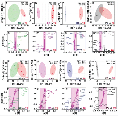
Table 3. Goodness-of-fit of the PLS-DA models obtained from 1D CPMG and 1D DE of rat serum samples.
Table 4. Key observed metabolic differences between the healthy control and DMH rat sera. chemical shift, variation, VIP score and p-values of the individual biomarkers are given. p-values less than 0.05 were considered as significant. The metabolic differences between DMH and DMH+5-FU, DMH and DMH+PA-10 mg, and DMH and DMH+PA-25 mg have also been presented.