1,027
Views
7
CrossRef citations to date
0
Altmetric
Bedside to Bench Report

Ovarian metastasis from lung adenocarcinoma with ALK-positive rearrangement detected by next generation sequencing: A case report and literatures review

, , , , &
Pages 279-284 | Received 06 Dec 2016, Accepted 19 Mar 2017, Published online: 20 Apr 2017

Figures & data

Figure 1. Shows the initial assessment before treatment by means of computed tomography (CT) scan of chest with contrast. The arrows are that: (A) left lower lobe masses (B) mediastinal lymphadenopathies (C) pleural metastasis.
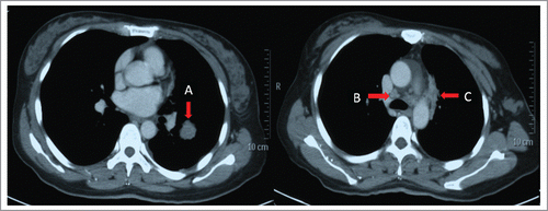
Figure 1. Shows the initial assessment before treatment by means of computed tomography (CT) scan of chest with contrast. The arrows are that: (A) left lower lobe masses (B) mediastinal lymphadenopathies (C) pleural metastasis.

Figure 2. The cytological examination revealed adenocarcinoma cells. Wright-Giemsa Stain (10 × 40) is used in both left and right panel.
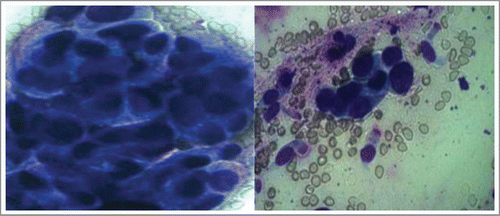
Figure 2. The cytological examination revealed adenocarcinoma cells. Wright-Giemsa Stain (10 × 40) is used in both left and right panel.

Figure 3. Shows the response at the different evaluation time. The panels mean that: (A) The response evaluation of primary tumor after 2 courses chemotherapy is partial response (PR). The arrow in panel A shows the residual mass after 2 courses first-line chemotherapy. (B) The response of primary tumor after 4 courses chemotherapy is complete response (CR). (C) The response of primary tumor after 6 courses chemotherapy is CR. (D) Response before the last maintenance therapy was evaluated and stable disease persists.
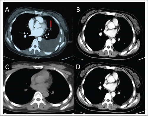
Figure 3. Shows the response at the different evaluation time. The panels mean that: (A) The response evaluation of primary tumor after 2 courses chemotherapy is partial response (PR). The arrow in panel A shows the residual mass after 2 courses first-line chemotherapy. (B) The response of primary tumor after 4 courses chemotherapy is complete response (CR). (C) The response of primary tumor after 6 courses chemotherapy is CR. (D) Response before the last maintenance therapy was evaluated and stable disease persists.

Figure 4. The panels means that: (A) CT scan showed left ovarian metastasis. The red arrow in panel A shows the ovarian metastatic mass, which diameter is about 10 cm. (B and C) There is no evidence of progression on regular follow-up examination.

Figure 4. The panels means that: (A) CT scan showed left ovarian metastasis. The red arrow in panel A shows the ovarian metastatic mass, which diameter is about 10 cm. (B and C) There is no evidence of progression on regular follow-up examination.

Figure 5. The panels means that: (A) The pathological diagnosis of the specimens was metastatic pulmonary adenocarcinoma. (H-E staining 10 × 40) (B) TTF-1 is positive in IHC staining. (C) Napsin A is positive in IHC staining.

Figure 5. The panels means that: (A) The pathological diagnosis of the specimens was metastatic pulmonary adenocarcinoma. (H-E staining 10 × 40) (B) TTF-1 is positive in IHC staining. (C) Napsin A is positive in IHC staining.

Table 1. Patients with metastasis in non-small cell lung cancer who harboring ALK rearrangement.

Reprints and Corporate Permissions

Please note: Selecting permissions does not provide access to the full text of the article, please see our help page How do I view content?

To request a reprint or corporate permissions for this article, please click on the relevant link below:

Academic Permissions

Please note: Selecting permissions does not provide access to the full text of the article, please see our help page How do I view content?

Obtain permissions instantly via Rightslink by clicking on the button below:

If you are unable to obtain permissions via Rightslink, please complete and submit this Permissions form. For more information, please visit our Permissions help page.