Figures & data
Table 1. Patient characteristics.
Table 2. Changes in WBC count and liver and kidney function after treatment.
Figure 1. Changes in the lymphocyte subsets before and after NK cell treatment (**P < 0.01).
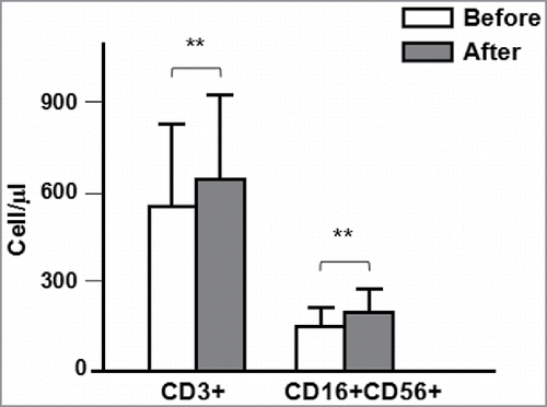
Table 3. Changes in the cytokine levels after treatment.
Figure 2. Changes in the TK1 level and number of CTCs before and after NK cell treatment (**P < 0.01).
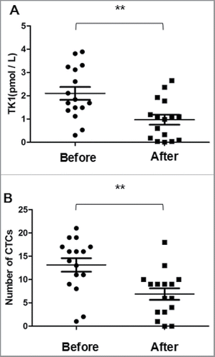
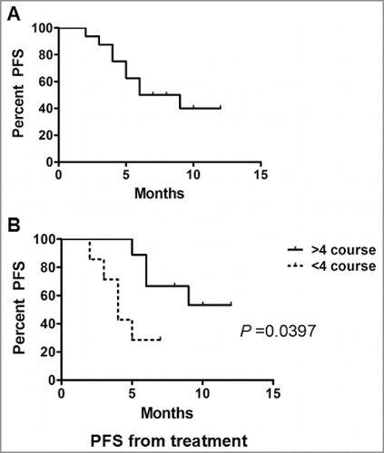
Table 4. Number of immunotherapy courses and clinical outcomes of NK cell immunotherapy at post-treatment 3 months.
Figure 3. CT images of a 52-year-old man with bilateral pulmonary metastasis who underwent 4 courses of HANK cell immunotherapy. The blue arrows in panels (A) and(B) indicate the tumor. The yellow arrows in panels (A) and (B) indicate pleural effusion. (A) CT scan showing a tumor of approximately 2 cm diameter with bilateral pleural effusion; (B) CT scan taken at 3 months after NK immunotherapy showing that the size of the tumor was 1.4 cm and the right-sided pleural effusion had disappeared.
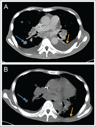
Figure 4. CT images of a 55-year-old man with bilateral pulmonary metastasis who underwent 4 courses of HANK cell immunotherapy. The circular area and blue arrows in panels (A) and (B) indicate the tumors. (A) Contrast-enhanced CT scan taken in the venous phase showing 2 lung multiple metastatic tumors (circular areas), with the larger tumor having a diameter of more than 0.5 cm (blue arrows). (B) Contrast-enhanced CT scan taken at 3 months after NK cell immunotherapy showing decrease in pulmonary metastasis (circular area) and a decrease in the diameter of the larger tumor to about 0.3 cm (blue arrows).
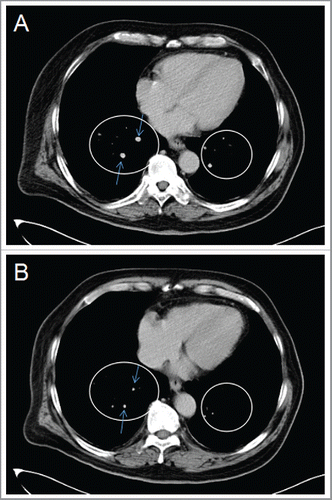
Figure 5. CT images of a 38-year-old man who underwent 6 courses of HANK cell immunotherapy for recurrent HCC. The blue arrows in panels (A) and (B) indicate the tumor. (A) Contrast-enhanced CT scan in the venous phase showing a tumor that was approximately 1.8 × 1.4 cm in size. (B) Contrast-enhanced CT scan taken 3 months after NK cells immunotherapy showing that the size of the tumor was 1.8 × 1.2 cm.
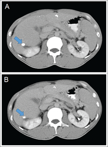
Figure 6. Effect of the number of infusions of allogeneic activated NK cells on progression-free survival. Progression-free survival from treatment of patients who received <4 infusions (a) and >4 infusions (b).