Figures & data
Table 1. Baseline characteristics of breast cancer patients included in this study (n = 57).
Figure 1. Representative examples of immunohistochemical expression of pMST1/2 and pLATS1/2 in 4 breast cancer cases (a,b,c,d). Panels a and b show 2 cases with cytoplasmic pMST1/2 (a) and cytoplasmic pLATS1/2 (b) expression with concomitant expression in stromal TILs. Panels c and d show 2 cases with nuclear pMST1/2 (c) and nuclear pLATS1/2 (d) expression with concomitant expression in stromal TILs. Slide magnification x 40, inset magnification x 20. Scale bar 30 μm.
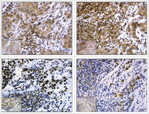
Table 2. expression pattern of pMST1/2 and pLATS1/2 in HER2-positive and triple-negative breast cancer (n = 57).
Table 3. Significant or borderline significant associations between the molecular markers of interest (pMST1/2 and pLATS1/2) and clinical-pathological factors.
Figure 2. Bar charts illustrating the association between pMST1/2cyt and TAZ (panel A), and between pMST1/2nuc and pChk1 (panel B).
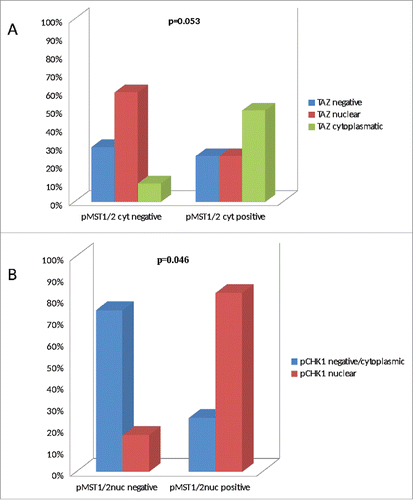
Table 4. Relationship between pMST1/2 expression and pCR (n=57).
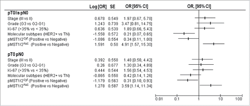
Figure 3. Univariate regression models for pCR (pT0/is pN0 and pT0 pN0) illustrating OR and 95%CI: clinical-molecular variables are reported including stage (III vs II), grade (G3 vs G1–2), Ki-67 levels (high vs low), molecular subtypes (HER2-positive vs triple-negative), pMST1/2cyt (positive vs negative) and pMST1/2nuc (positive vs negative).