Figures & data
Figure 1. The 4 steps of immune surveillance in black boxes; factors that enhance the efficacy and contribute to an effective immune surveillance in green boxes; factors that impair an effective immune surveillance and help melanoma escape immune attack in red boxes. DC: dendritic cells, TVEC: Talimogene laherparepvec, IFN-y: interferon gamma, CCL4: chemokine CCL4, TAP: Transporter associated with antigen processing, PD1: programmed death receptor 1, CTLA4: cytotoxic lymphocyte antigen 4, LAG3: lymphocyte-activation gene-3, TIM3: T-cell immunoglobulin mucin-3, BTLA: B- and T-lymphocyte attenuator, FLIP: FLICE-inhibitory proteins, IAPs: inhibitor of apoptosis proteins, CD95: death receptor also known as Fas, TRAIL/Apo2L: TNF-related apoptosis-inducing ligand, DISC: death-inducing signaling complex.
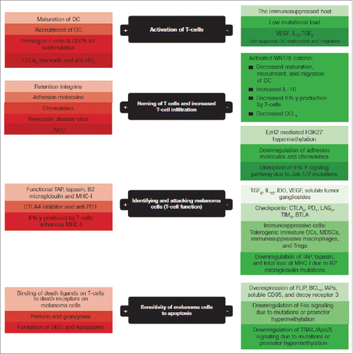
Figure 2. Phagocytosis of tumor antigens by dendritic cells (DCs) and the antigen-presenting machinery that helps load the tumor peptides onto MHC-II molecules on the DC surface; 1) Endocytosis of tumor specific antigen (TSA), 2) Formation of endocytic vesicle that transports the tumor specific antigens into the cytosol, 3) the tumor specific antigens are degraded in the cytosol by proteasomes that degrades the proteins into peptides, 4) The tumor peptides are transported to the endoplasmic reticulum (ER) by transporter associated with antigen processing (TAP), 5) In the ER the tumor peptides are loaded on the MHC I or MHC II molecules to form complexes that (6) that moves through the Golgi apparatus and got displayed on the dendritic cell wall where they interact with the T cell receptors.
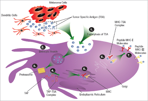
Figure 3. T cell activation and the mechanism of actions of both PD1 and CTLA4 inhibitors; PD1: programmed death 1 receptor, CTLA4: cytotoxic lymphocyte antigen-4, MHC: major histocompatibility complex, TCR: T cell receptor, TSA: tumor specific antigen, APS: antigen presenting cell, PDL1: programmed death ligand-1.
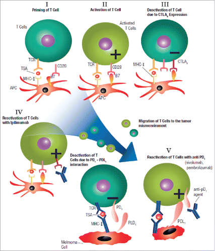
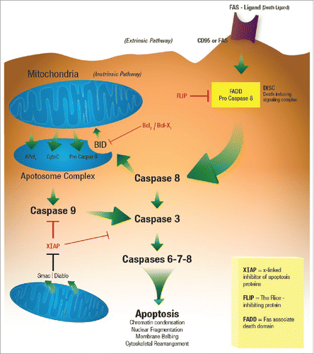
Figure 4. Apoptosis.