Figures & data
Figure 1. Transmission electron microscopy. ZnO-NPs typically clotted together in various intracellular patches of different amounts (indicated by black arrows). Scale bar represents 250 nm, image was taken at 40.000x magnification.
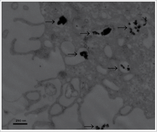
Figure 2. Immunofluorescence microscopy of the EGF-Receptor. Regular staining of EGFR on the cell membrane without Zn-NPs (A) as well as after addition of Zn-NPs (B), showing no significant difference.
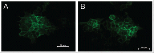
Figure 3. MTT-assay for FaDu cell line. Time points were 24 hours (A), 48 hours (B) and 72 hours (C). At 48 and 72 hours, ZnO-NPs increased tumor cell survival at 1 µM Cetuximab, whereas no significant influence was found at 0.1 µM and 10 µM Cetuximab or after 24 hours of incubation.
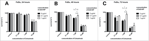
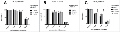
Figure 4. MTT-assay for HLaC cell line. Time points were 24 hours (A), 48 hours (B) and 72 hours (C). At 72 hours, ZnO-NPs increased tumor cell survival at 1 µM Cetuximab, whereas no significant influence was found at 0.1 µM and 10 µM Cetuximab or after 24 and 48 hours of incubation.