Figures & data
Figure 1. HSP27 was upregulated in PHC patient's samples and cell lines. (A) The expression of Hsp27 protein in healthy control group (Control) group, BLL group and PHC group via immunohistochemistry assay. Magnification, 200 ×. (B) The mRNA level of Hsp27 in healthy control group (Control), benign liver lesions patients group (BLL) and primary hepatocellular carcinoma patients group (PHC) via real time RT-PCR assay. Fold change of HSP27 mRNA in BLL and PHC group was compared with Control group. (C) The mRNA level of Hsp27 in L02 cells, Huh7 cells and HepG2 cells via real time RT-PCR assay. Fold change of HSP27 mRNA in Huh7 and HepG2 cells was compared with L02 cells. (D) The expression of HSP27 protein in L02 cells, Huh7 cells and HepG2 cells via western blot assay. The ratios of HSP27 to GAPDH for 3 independent experiments are shown as indicated below the blots.
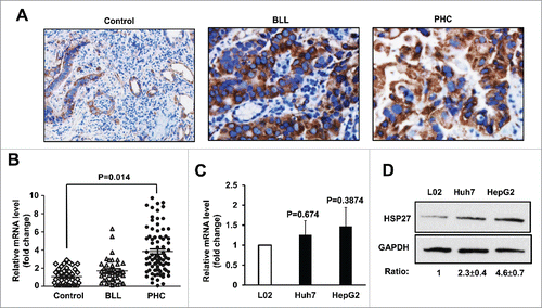
Table 1. Relationship between plasma HSP27 level and clinicopathological features in patients with HCC.
Figure 2. HSP27 siRNA reduces the proliferation and invasion of the HCC cells. (A) The expression of HSP27 protein in Huh7 cells and HepG2 cells transfected with Scramble siRNA (Scr) or siRNA targeting Hsp27 (siRNA) for 48 hours via western blot assay, respectively. GAPDH was used as the loading control for each group. The ratios of HSP27 to GAPDH for 3 independent experiments are shown as indicated below the blots. (B) The relative cell growth rate (normalized with the cell numbers at day 0) in Huh7 cells (left panel) and HepG2 cells (right panel) transfected with Scr or siRNA for 0, 24, 48, 72 and 96 hours after transfection by adding 10 μl of CCK8 reagent. *, p < 0.05 and **, p < 0.01 in siRNA group compared with Scr group. (C) The invasion capability in Huh7 cells and HepG2 cells transfected with Scr or siRNA via transwell assay after 48 hours. (D) Quantitative analysis of the invasion cell numbers per well of a 24-well plate in the Huh7 and HepG2 cells transfected with scramble control or si-HSP27. Data presents Mean±SD for 12 independent visions per group in the Huh7 and HepG2 cells, respectively. *, p < 0.05, **, p < 0.01, the scramble groups vs. si-HSP27 group.
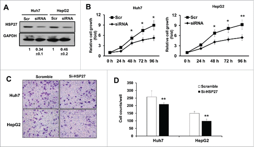
Figure 3. HSP27 was post-transcriptionally modified by protein degradation. (A) The expression of HSP27 protein in L02 cells, Huh7 cells and HepG2 cells treated with MG132 (20 μM) at 0, 2, 4, 6 hours via western blot assay. The ratios of HSP27 to GAPDH for 3 independent experiments are shown as indicated below the blots. (B) The expression of SUMO2/3 protein in L02 cells, Huh7 cells and HepG2 cells via western blot assay. The ratios of SUMO2/3 to GAPDH for 3 independent experiments are shown as indicated below the blots. (C) The correlation analysis of the protein expression between HSP27 and SUMO2/3. (D) Immunofluorescence of nucleus (DAPI, blue), SUMO2/3 (green) and HSP27 (red) in Huh7 cells and HepG2 cells.
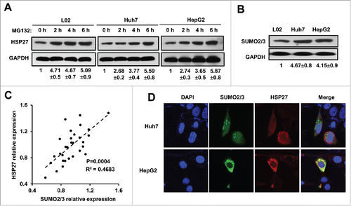
Figure 4. SUMO 2/3 directly binds HSP27 protein. (A) Shown is the amino acid sequence of HSP27 protein and the binding site of SUMO2/3 (bold and underlined fonts). Immunoprecipitation assay showed the interaction of SUMO2/3 with HSP27 in (B) Huh7 cells and (C) HepG2 cells. 20 μg of the whole cell lysate was served as the input control, and 2 μg of IgG control or α-HSP27 monoclonal antibody was used to precipitate the target protein HSP27, and after blotting, the SUMO2/3 antibody was incubated to detect the binding on HSP27 protein at dilution of 1:500. The ratios of SUMO2/3 in the IgG or α-HSP27 group to that in the input group for 3 independent experiments are shown as indicated below the blots.
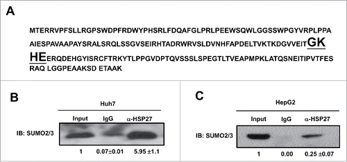
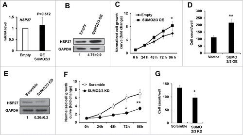
Figure 5. SUMO2/3 promotes PHC cell proliferation and invasion. (A) The mRNA level of HSP27 in Huh7 cells transfected with empty vector (Empty) or SUMO2/3 expressing plasmid for 48 hours was detected using real time PCR. (B) Shown are western blot assays to determine the expression of HSP27 protein in Huh7 cells transfected with empty vector control or SUMO2/3 overexpression plasmids for 48 hours. The ratios of HSP27 to GAPDH for 3 independent experiments are shown as indicated below the blots. (C) The relative cell growth rate in Huh7 cells transfected with SUMO2/3 overexpression plasmid or Empty vector via cck-8 assay. Cell growth has been normalized to the day 0 and shown as fold change. (D) The invasion cell numbers per well of a 24-well plate of Huh7 cells transfected with SUMO2/3 overexpression plasmid or Empty vector for 48 hours via transwell assay. (E) Shown are western blot assays to determine the expression of HSP27 protein in Huh7 cells transfected with scramble control or siRNA specially targeting SUMO2/3 for 48 hours. The ratios of HSP27 to GAPDH for 3 independent experiments are shown as indicated below the blots. (F) The relative cell growth rate in Huh7 cells transfected with scramble control or siRNA specially targeting SUMO2/3 via cck-8 assay. Cell growth has been normalized to the day 0 and shown as fold change. (G) The invasion cell numbers per well of a 24-well plate of Huh7 cells transfected with scramble control or siRNA specially targeting SUMO2/3 for 48 hours via transwell assay. *, p < 0.05 and **, p < 0.01; vector control vs. OE or KD vectors.