Figures & data
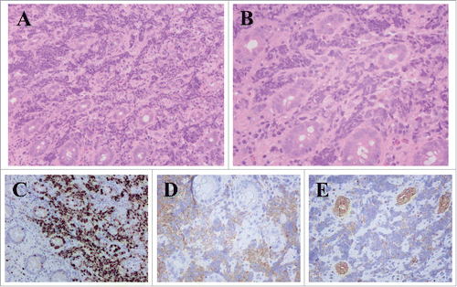
Figure 1. Pathological images from esophageal SCC. A. Hematoxylin-eosin stain (× 100). B. Hematoxylin-eosin stain (× 200). C.D.E. Immunohistochemical staining in esophageal SCC (× 200). Ki-67: 40%, Syn (+), CD56 (+).
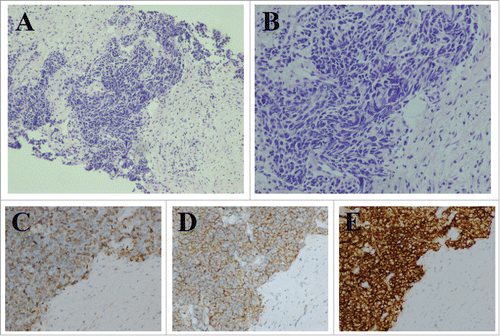
Figure 2. CT scan of abdomen demonstrating a swollen appendix with an unclear border, uneven density, and heterogeneous enhancement is visible (arrow).
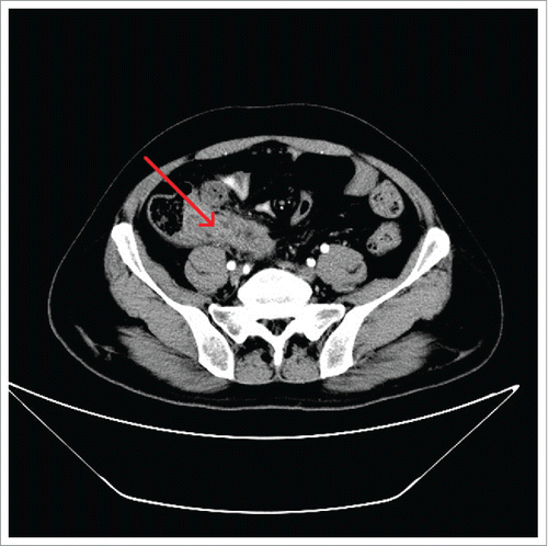
Figure 3. Pathological results from SCC of appendix. A. Hematoxylin-eosin stain (× 100). B. Hematoxylin-eosin stain (× 200). C.D.E. Immunohistochemical staining in SCC of appendix. Ki-67: 80% (× 100), Syn (+) (× 200), panCK (+) (× 200).