1,274
Views
27
CrossRef citations to date
0
Altmetric
Research Paper

Microvesicles releasing by oral cancer cells enhance endothelial cell angiogenesis via Shh/RhoA signaling pathway

, , , , &
Pages 783-791 | Received 01 Mar 2017, Accepted 24 Aug 2017, Published online: 13 Oct 2017

Figures & data

Figure 1. Immunohistochemical detection of Shh, Gli1 in OSCC specimens. A, cytoplasm expression of OSCC specimen for Shh (a1, low staining; a2, moderate staining; a3, strong staining); B, nuclear and cytoplasm expression of OSCC specimen for Gli1 (b1, low staining; b2, moderate staining; b3, strong staining). Bars indicate 100 um.
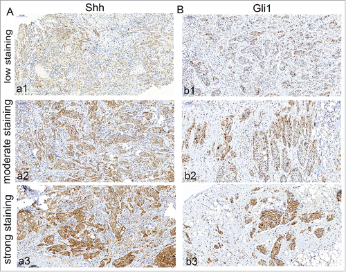
Figure 1. Immunohistochemical detection of Shh, Gli1 in OSCC specimens. A, cytoplasm expression of OSCC specimen for Shh (a1, low staining; a2, moderate staining; a3, strong staining); B, nuclear and cytoplasm expression of OSCC specimen for Gli1 (b1, low staining; b2, moderate staining; b3, strong staining). Bars indicate 100 um.

Figure 2. Immunohistochemical detection of Shh, Gli-1 in metastatic lymphnodes of OSCC specimens. A, cytoplasm of expression of metastatic lymphnodes for Shh (strong staining); B, nuclear and cytoplasm expression of metastatic lymphnodes for Gli1 (strong staining). Bars indicate 100 um.
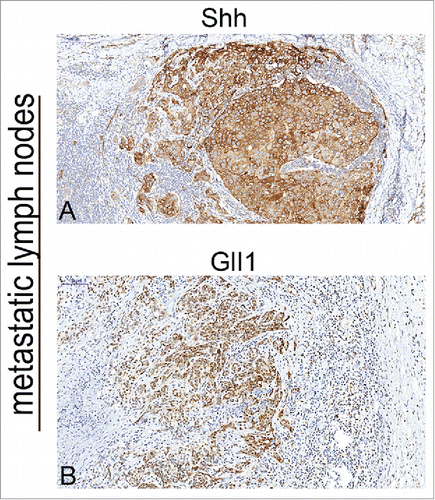
Figure 2. Immunohistochemical detection of Shh, Gli-1 in metastatic lymphnodes of OSCC specimens. A, cytoplasm of expression of metastatic lymphnodes for Shh (strong staining); B, nuclear and cytoplasm expression of metastatic lymphnodes for Gli1 (strong staining). Bars indicate 100 um.

Figure 3. The correlation between MVD and Shh expression. Microvesseldentisty in OSCC specimens. A, the representative picture showed the value of MVD >57, and the MVD was high in 37 of 80 OSCC specimens; B, the representative picture showed the value of MVD <57, and the MVD was low in 43 of 80 OSCC specimens; C, Correlation between Shh and MVD expression. Spearman correlation and linear regression were used to determine the relationship between Shh and MVD. MVD correlated positively with Shh expression (P < 0.01).
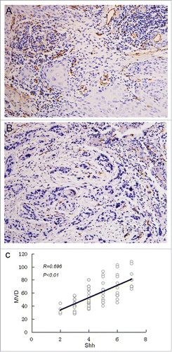
Figure 3. The correlation between MVD and Shh expression. Microvesseldentisty in OSCC specimens. A, the representative picture showed the value of MVD >57, and the MVD was high in 37 of 80 OSCC specimens; B, the representative picture showed the value of MVD <57, and the MVD was low in 43 of 80 OSCC specimens; C, Correlation between Shh and MVD expression. Spearman correlation and linear regression were used to determine the relationship between Shh and MVD. MVD correlated positively with Shh expression (P < 0.01).

Table 1. The correlation between Shh and Gli1 expression.

Table 2. Correlation between Shh, Gli1, MVD expression and clinicopathologic factors.

Figure 4. The expression of Shh in OSCC cell lines, Cal27 derived MVs A, the expression of Shh protein was detected in OSCC cell lines tested by western blot; B, western blot was used to detect the expression of Shh in Cal27 lysates and Cal27-derived MVs. A difference of P< 0.05 was considered significant.
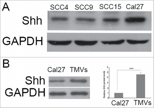
Figure 4. The expression of Shh in OSCC cell lines, Cal27 derived MVs A, the expression of Shh protein was detected in OSCC cell lines tested by western blot; B, western blot was used to detect the expression of Shh in Cal27 lysates and Cal27-derived MVs. A difference of P< 0.05 was considered significant.

Figure 5. The efficacy of Shh/RhoA pathway on tube formation of HUVECs in vitro. A, Phase-contrast micrographs showed that MVs harboring Shh enhanced network formation of HUVECs on Matrigel. The efficacy of MVs-enhanced network formation was inhibited by C3 transferase (5 ug/ml). The Shh (5 ug/ml) was used as the positive control. The treatment condition and the actual number of branch points ± SEM were underneath the image. Branch points were used to quantify angiogenesis; B, Shh associated tube formation was related with RhoA activation; C, the expression of Gli1 in HUVECs after rh-shh (5 ug/ml) treatment. A difference of P < 0.05 was considered significant.
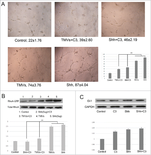
Figure 5. The efficacy of Shh/RhoA pathway on tube formation of HUVECs in vitro. A, Phase-contrast micrographs showed that MVs harboring Shh enhanced network formation of HUVECs on Matrigel. The efficacy of MVs-enhanced network formation was inhibited by C3 transferase (5 ug/ml). The Shh (5 ug/ml) was used as the positive control. The treatment condition and the actual number of branch points ± SEM were underneath the image. Branch points were used to quantify angiogenesis; B, Shh associated tube formation was related with RhoA activation; C, the expression of Gli1 in HUVECs after rh-shh (5 ug/ml) treatment. A difference of P < 0.05 was considered significant.

Figure 6. The effect of knockdown of RhoA on tube formation of HUVECs in vitro. A, The phase-contrast micrographs showed that TMVs enhanced network formation of HUVECs on Matrigel. The effect of TMVs-enhanced angiogenesis was inhibited by silencing the RhoA expression. The treatment condition and the actual number of branch points ± SEM were underneath the image. Branch points were used to quantify angiogenesis. B, As the histogram shown, the role of knockdown of RhoA in TMVs-mediated angiogenesis was similar with the data using C3. The western blot bands inserted in histogram showed the effect of silencing of RhoA. A difference of P < 0.05 was considered significant.
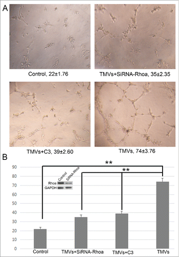
Figure 6. The effect of knockdown of RhoA on tube formation of HUVECs in vitro. A, The phase-contrast micrographs showed that TMVs enhanced network formation of HUVECs on Matrigel. The effect of TMVs-enhanced angiogenesis was inhibited by silencing the RhoA expression. The treatment condition and the actual number of branch points ± SEM were underneath the image. Branch points were used to quantify angiogenesis. B, As the histogram shown, the role of knockdown of RhoA in TMVs-mediated angiogenesis was similar with the data using C3. The western blot bands inserted in histogram showed the effect of silencing of RhoA. A difference of P < 0.05 was considered significant.

Reprints and Corporate Permissions

Please note: Selecting permissions does not provide access to the full text of the article, please see our help page How do I view content?

To request a reprint or corporate permissions for this article, please click on the relevant link below:

Academic Permissions

Please note: Selecting permissions does not provide access to the full text of the article, please see our help page How do I view content?

Obtain permissions instantly via Rightslink by clicking on the button below:

If you are unable to obtain permissions via Rightslink, please complete and submit this Permissions form. For more information, please visit our Permissions help page.