Figures & data
Figure 1. CT Chest with Contrast Demonstrating Right Hilar Disease with Right-sided Pleural Involvement (arrows).
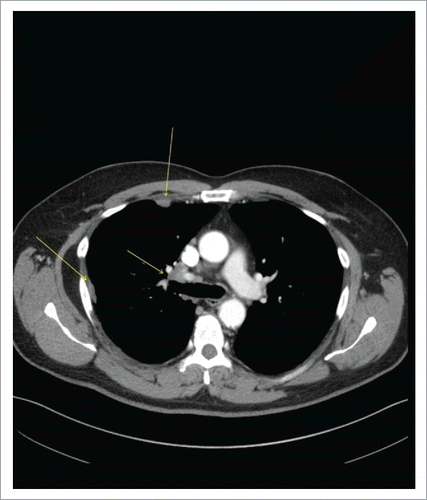
Figure 2. Histopathology and Immunohistochemical (IHC) Staining of Sequentially-Occurred NSCLC (ADC) and SCLC. Panel A: Histopathology and immunostaining of ADC of lung (Lymph node aspiration cytology). (a). H and E staining showing scattered enlarged cells with vesicular chromatin, visible nucleoli and moderately fine vacuolated cytoplasm. (b). IHC staining showing positive cytoplasmic stain for MOC31 (MOC31 is an immunological marker used for differentiation between metastatic ADC and lung mesothelioma where ADC is positive and mesothelioma is negative for MOC31). (c). IHC staining showing positive staining for CD15 (CD15 is a carbohydrate antigen on cells and can help during the confirmation process of lung ADC by differentiating it from mesothelioma which is negative for CD15 immunological staining). Panel B: Histopathology and immunostaining of transformed SCLC (Liver Biopsy). (a). H and E staining showing a diffuse infiltrate of polygonal neoplastic cells with a high nucleus to cytoplasm ratio, hyperchromatic nuclei, fine chromatin, inconspicuous nucleoli and focally forming vague pseudorosettes. (b). IHC staining showing positive cytoplasmic staining for synaptophysin. (c). IHC staining showing positive staining for CD56. (d). IHC staining showing very weak patchy staining for P63 (P63 is a nuclear marker used for immunostaining to differentiate squamous cell CA from ADC and is very strongly positive for SCC in contrast to ADC of the lung).
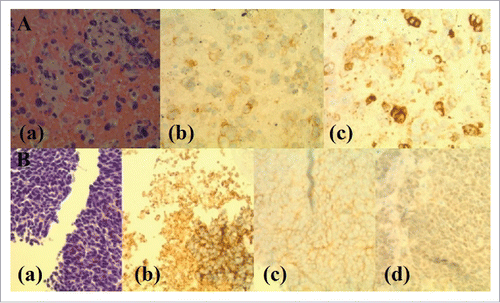
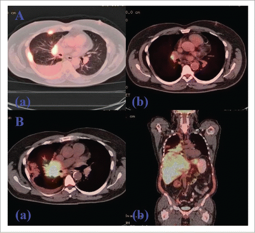
Figure 3. PET/CT of Chest Showing Initial Response with Subsequent Progression Following TKI-Exposure (afatinib). (A). PET/CT showing interval response after 4 months treatment of afatinib. (a). FDG-avid thoracic LAD and right pleural disease before the initiation of afatinib. (b). Marked improvement of lung cancer on PET/CT showing a solitary residual right hilar LN after four months of TKI treatment. (B). PET/CT showing progression after 10 months treatment of afatinib. (a). Axial view showing a marked progression of metastatic lung disease with diffuse hepatic and skeletal involvement along with the progression of right perihilar mass. (b). Coronal view showing the same progression following post-TKI resistance.