
Figures & data
Table 1. Patient characteristics.
Figure 1. A 63-year-old man with hepatocellular carcinoma. Preoperative MR and CEUS images showed a mass of 2.2#1.7 cm in size in the caudate lobe (a, b and c) (white arrows). EUS suggested laser fiber inserted into the tumor (d) and then total enhancement of the lesion (e) (white arrows). One year later, substance phase MR image showed that the mass has a complete response (f) (white arrows).
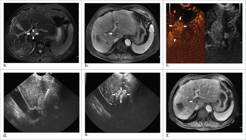
Figure 2. Representative images from a 70-year-old man diagnosed liver metastasis from colon cancer. T1 and T2 MR images revealed a tumor about 2.1#1.7 cm in the caudate lobe (a and b) (white arrows). One laser fiber was ablating the tumor with local enhancement (c) (white arrows). T2 MR image two months obtained after ablation showed complete response in the tumor (d) (white arrows). At the corresponding ultrasounography, it also showed a complete necrosis without any enhanced perfusion in CEUS (e and f) (white arrows).
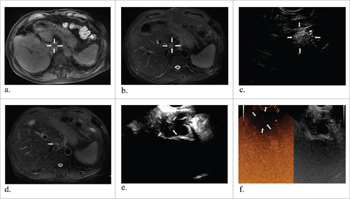
Figure 3. A 57-year-old man with hepatocellular carcinoma. MR obtained one month before ablation showed the tumor measuring 1.3#1.2 cm in diameter in the caudate lobe (a) (arrowheads). Preoperative EUS indicated a low echo area (b) (arrowheads) and it had increased echogenicity covering the whole mass after ablation (c) (arrowheads). After one month, enhanced MR revealed the lesion complete necrosis (d) (arrowheads).
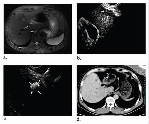
Figure 4. Representative images from a 54-year-old man diagnosed with hepatocellular carcinoma. Preoperative T2 MR image shows a round tumor about 1.1#1.0 cm in the left liver (a) (arrowheads). After enhanced perfusion of this tumor under CEUS guidance (b) (arrowheads), then a laser fiber ablated the target tumor (c) (arrowheads), and immediately CEUS showed no enhanced perfusion (d) (arrowheads). During six-month follow-up after ablation, T1 (e) and substance phase MR images (f) showed the mass was successfully ablated (arrowheads).
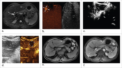
Table 2. The detailed description of EUS-guided laser ablation for tumors.
Table 3. Summary of EUS-guided interventional treatment of tumors in 4 published literatures.