1,370
Views
1
CrossRef citations to date
0
Altmetric
Research Paper

LncRNA ADPGK-AS1 promotes pancreatic cancer progression through activating ZEB1-mediated epithelial–mesenchymal transition

, , , , , & show all
Pages 573-583 | Received 18 Aug 2017, Accepted 29 Dec 2017, Published online: 19 Apr 2018

Figures & data

Table 1. Primers for qRT-PCR.

Figure 1. LncRNA and miRNA expression profiles in pancreatic cancer (A) Volcano plot of LncRNA expression profile in PC; (B) Heatmap of LncRNA expression profile in PC; (C) Volcano plot of miRNA expression profile in PC; (D) Heatmap of lncRNA expression profile in PC; (E) The lncRNA/miRNA interaction, predicted an direct association between miR-205-5p and ADPGK-AS1.
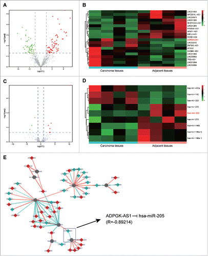
Figure 1. LncRNA and miRNA expression profiles in pancreatic cancer (A) Volcano plot of LncRNA expression profile in PC; (B) Heatmap of LncRNA expression profile in PC; (C) Volcano plot of miRNA expression profile in PC; (D) Heatmap of lncRNA expression profile in PC; (E) The lncRNA/miRNA interaction, predicted an direct association between miR-205-5p and ADPGK-AS1.

Figure 2. LncRNA ADPGK-AS1 directly targeted miR-205-5p (A) Relative expression level of ADPGK-AS1 in PANC-1 and SW-1990 cells; (B) Relative expression level of miR-205-5p in PANC-1 and SW-1990 cells; (C) The predicted binding site between miR-205-5p and ADPGK-AS1; (D) The relative luciferase activity of ADPGK-AS1+miR-205-5p was significantly lower; (E) miR-205-5p expression could be promoted by miR-205-5p mimics transfection while suppressed by ADPGK-AS1 transfection; (F) ADPGK-AS1 expression could be promoted by ADPGK-AS1 transfection while miR-205-5p mimics transfection had little effect on ADPGK-AS1 expression. (*P < 0.05, **P < 0.01, compared with control group).
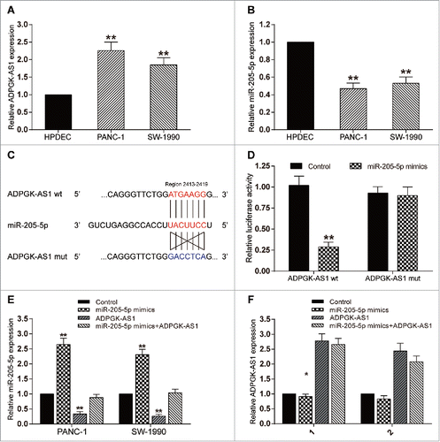
Figure 2. LncRNA ADPGK-AS1 directly targeted miR-205-5p (A) Relative expression level of ADPGK-AS1 in PANC-1 and SW-1990 cells; (B) Relative expression level of miR-205-5p in PANC-1 and SW-1990 cells; (C) The predicted binding site between miR-205-5p and ADPGK-AS1; (D) The relative luciferase activity of ADPGK-AS1+miR-205-5p was significantly lower; (E) miR-205-5p expression could be promoted by miR-205-5p mimics transfection while suppressed by ADPGK-AS1 transfection; (F) ADPGK-AS1 expression could be promoted by ADPGK-AS1 transfection while miR-205-5p mimics transfection had little effect on ADPGK-AS1 expression. (*P < 0.05, **P < 0.01, compared with control group).

Figure 3. LncRNA ADPGK-AS1 induced proliferation, decrease of apoptosis rate, migration, and invasion of PC cells (A) CCK-8: PANC-1/SW-1990 cell viability could be promoted by ADPGK-AS1 while suppressed by miR-205-5p mimics; (B) TUNEL: PANC-1/SW-1990 cell apoptosis could be reduced by ADPGK-AS1 while enhanced by miR-205-5p mimics; (C) Transwell: PANC-1/SW-1990 cell migration could be promoted by ADPGK-AS1 while suppressed by miR-205-5p mimics; (D) Transwell: PANC-1/SW-1990 cell invasion could be promoted by ADPGK-AS1 while suppressed by miR-205-5p mimics. (*P < 0.05, **P < 0.01, compared with control group).
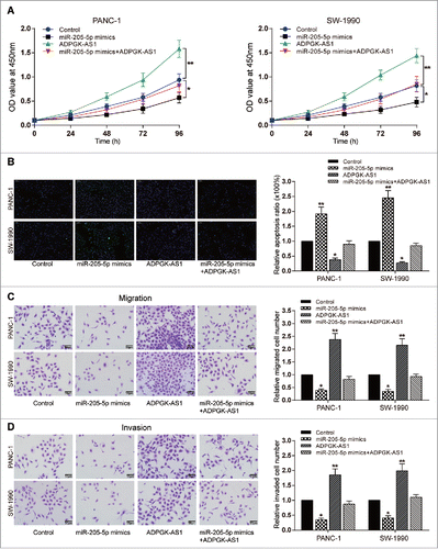
Figure 3. LncRNA ADPGK-AS1 induced proliferation, decrease of apoptosis rate, migration, and invasion of PC cells (A) CCK-8: PANC-1/SW-1990 cell viability could be promoted by ADPGK-AS1 while suppressed by miR-205-5p mimics; (B) TUNEL: PANC-1/SW-1990 cell apoptosis could be reduced by ADPGK-AS1 while enhanced by miR-205-5p mimics; (C) Transwell: PANC-1/SW-1990 cell migration could be promoted by ADPGK-AS1 while suppressed by miR-205-5p mimics; (D) Transwell: PANC-1/SW-1990 cell invasion could be promoted by ADPGK-AS1 while suppressed by miR-205-5p mimics. (*P < 0.05, **P < 0.01, compared with control group).

Figure 4. MiR-205-5p directly targeted ZEB1 (A-B) Western blot: in PANC-1 and SW-1990 cells, E-cadherin expression was promoted by miR-205-5p mimics while suppressed by miR-205-5p inhibitor; ZEB1 and N-cadherin expression was suppressed by miR-205-5p mimics while promoted by miR-205-5p inhibitor; (C) The predicted binding site between miR-205-5p and ZEB1;(D) The relative luciferase activity of ZEB1 3′UTR wt+miR-205-5p mimics was remarkably lower; (E) Western blot: in PANC-1 and SW-1990 cells, ZEB1 expression was promoted by ZEB1 cDNA while suppressed by ZEB1 shRNA. (*P < 0.05, **P < 0.01, compared with control group).
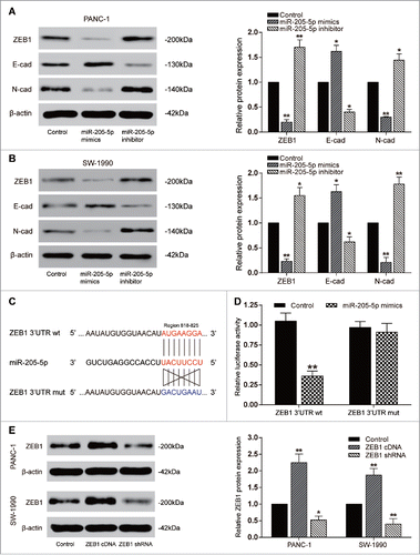
Figure 4. MiR-205-5p directly targeted ZEB1 (A-B) Western blot: in PANC-1 and SW-1990 cells, E-cadherin expression was promoted by miR-205-5p mimics while suppressed by miR-205-5p inhibitor; ZEB1 and N-cadherin expression was suppressed by miR-205-5p mimics while promoted by miR-205-5p inhibitor; (C) The predicted binding site between miR-205-5p and ZEB1;(D) The relative luciferase activity of ZEB1 3′UTR wt+miR-205-5p mimics was remarkably lower; (E) Western blot: in PANC-1 and SW-1990 cells, ZEB1 expression was promoted by ZEB1 cDNA while suppressed by ZEB1 shRNA. (*P < 0.05, **P < 0.01, compared with control group).
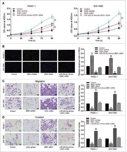
Figure 5. MiR-205-5p reversed ZEB1-induced proliferation, decrease of apoptosis rate, migration, and invasion of PC cells (A) CCK-8: PANC-1/SW-1990 cell viability could be promoted by ZEB1 cDNA while suppressed by ZEB1 shRNA; (B) TUNEL: PANC-1/SW-1990 cell apoptosis could be reduced by ZEB1 cDNA while enhanced by ZEB1 shRNA; (C) Transwell: PANC-1/SW-1990 cell migration could be promoted by ZEB1 cDNA while suppressed by ZEB1 shRNA; (D) Transwell: PANC-1/SW-1990 cell invasion could be promoted by ZEB1 cDNA while suppressed by ZEB1 shRNA. (*P < 0.05, **P < 0.01, compared with control group).

Figure 5. MiR-205-5p reversed ZEB1-induced proliferation, decrease of apoptosis rate, migration, and invasion of PC cells (A) CCK-8: PANC-1/SW-1990 cell viability could be promoted by ZEB1 cDNA while suppressed by ZEB1 shRNA; (B) TUNEL: PANC-1/SW-1990 cell apoptosis could be reduced by ZEB1 cDNA while enhanced by ZEB1 shRNA; (C) Transwell: PANC-1/SW-1990 cell migration could be promoted by ZEB1 cDNA while suppressed by ZEB1 shRNA; (D) Transwell: PANC-1/SW-1990 cell invasion could be promoted by ZEB1 cDNA while suppressed by ZEB1 shRNA. (*P < 0.05, **P < 0.01, compared with control group).

Figure 6. ADPGK-AS1/miR-205-5P regulated PC tumor growth through EMT in vivo (A) Images of the excised tumors at 30th day were presented; (B) Tumor volume of miR-205-5p group was significantly lower while which of ADPGK-AS1 group was significantly higher; (C) Tumor weight of miR-205-5p group was significantly lower while which of ADPGK-AS1 group was significantly higher; (D) Total RNA in tumor tissues was extracted and qRT-PCR was utilized to measure the expression of ADPGK-AS1 and miR-205-5p. (E) Immunohistochemistry staining of Ki-67 was performed to value the cell viability during tumorigenesis. (F) Western blot: In tumor tissues, E-cadherin expression was promoted by miR-205-5p while suppressed by ADPGK-AS1; ZEB1 and N-cadherin expressions were suppressed by miR-205-5p while promoted by ADPGK-AS1. (*P < 0.05, **P < 0.01, compared with control group).
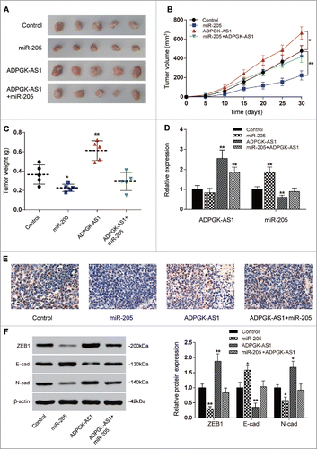
Figure 6. ADPGK-AS1/miR-205-5P regulated PC tumor growth through EMT in vivo (A) Images of the excised tumors at 30th day were presented; (B) Tumor volume of miR-205-5p group was significantly lower while which of ADPGK-AS1 group was significantly higher; (C) Tumor weight of miR-205-5p group was significantly lower while which of ADPGK-AS1 group was significantly higher; (D) Total RNA in tumor tissues was extracted and qRT-PCR was utilized to measure the expression of ADPGK-AS1 and miR-205-5p. (E) Immunohistochemistry staining of Ki-67 was performed to value the cell viability during tumorigenesis. (F) Western blot: In tumor tissues, E-cadherin expression was promoted by miR-205-5p while suppressed by ADPGK-AS1; ZEB1 and N-cadherin expressions were suppressed by miR-205-5p while promoted by ADPGK-AS1. (*P < 0.05, **P < 0.01, compared with control group).

Reprints and Corporate Permissions

Please note: Selecting permissions does not provide access to the full text of the article, please see our help page How do I view content?

To request a reprint or corporate permissions for this article, please click on the relevant link below:

Academic Permissions

Please note: Selecting permissions does not provide access to the full text of the article, please see our help page How do I view content?

Obtain permissions instantly via Rightslink by clicking on the button below:

If you are unable to obtain permissions via Rightslink, please complete and submit this Permissions form. For more information, please visit our Permissions help page.