
Figures & data
Table 1. Demographic and laboratory data of all patients and controls.
Table 2. The qPCR expression levels of the serum microRNAs in the studied groups.
Figure 1. Box plot diagrams showing the expression of (A) miR-16, (B) miR-34a, (C) miR-125a, (D) miR-139, (E) miR-145, (F) miR-199a, and (G) miR-221 in Hepatitis C virus (HCV)-induced hepatocellular carcinoma (HCC) patients. The box indicates the 25th and 75th percentile of the data and the middle line indicates the median. A line extends from the minimum to the maximum value, excluding outliers that are displayed as separate points.
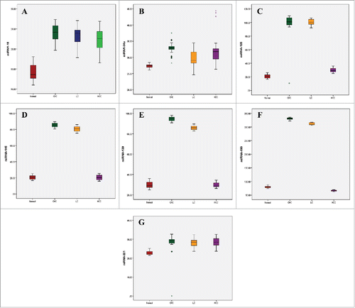
Table 3. Area Under the Curve (AUC), Confidence Interval (CI), and p-values for the circulating miRNAs.
Table 4. The sensitivity and specificity of the microRNAs.