
Figures & data
Figure 1. Immunohistochemical staining (IHC) of DLBCL tissue sample. IHC staining of CD5 (A). (Original magnification, *100). Positive IHC staining of CD5 (B), CD20 (C), CD79a (D), MUM1 (G), and negative staining of CD10 (E), Bcl-6 (F), MYC (H), CD3 (I). (Original magnification, *400).
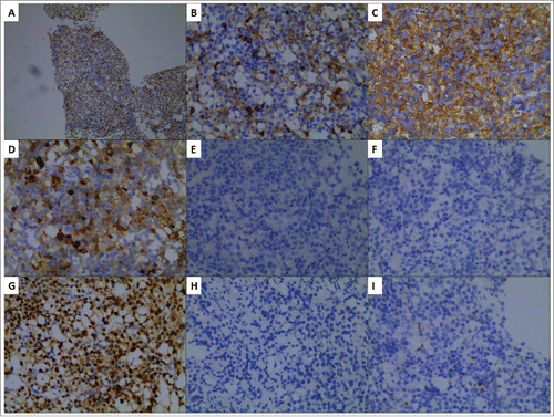
Figure 2. PET/CT scan before the start of treatment (A) and after three cycles of R2-GDP (B). The yellow arrows demonstrated the lesions where abdominal masses disappeared (B).
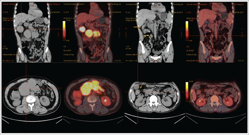
Table 1. Outcomes of CD5+ DLBCL treated with rituximab containing chemotherapy.