Figures & data
Figure 1. LncRNA PTCSC3 was low-expressed in ATC tissues. (A) The expression of PTCSC3 in FTC (n = 20) and ATC tissues (n = 20) was quantified by qRT-PCR. **P < 0.01 vs. FTC; the expression of proteins was analyzed by western blot. (B) The positive expression rate of CD133 was analyzed by flow cytometry. **P < 0.01 vs. FTC; the levels of ALDH and MDR-1 were analyzed by western blot.
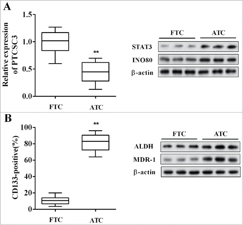
Figure 2. LncRNA PTCSC3 was low-expressed in ATC cells. (A) The expression of PTCSC3 in FTC 238, FTC 133 and 8505C cells was quantified by qRT-PCR. **P < 0.01 vs. FTC 238; the expression of proteins was analyzed by western blot. (B) The positive expression rate of CD133 in FTC 238, FTC 133 and 8505C cells was analyzed by flow cytometry. **P < 0.01 vs. FTC 238; the levels of ALDH and MDR-1 were analyzed by western blot.
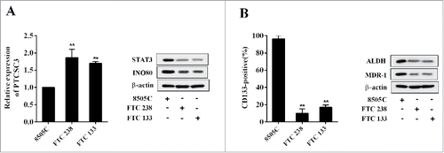
Figure 3. Over-expressed PTCSC3 inhibited the drug resistance of ATC. (A) The cytotoxicity of DOX to FTC 238 and FTC 133 was detected by MTT assay. **P < 0.01 vs. si-control. (B) The cytotoxicity of DOX to 8505C was detected by MTT assay. **P < 0.01 vs. pcDNA.
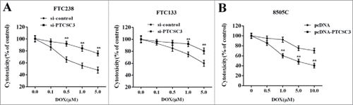
Figure 4. LncRNA PTCSC3 negatively regulated STAT3. (A) Combination condition between PTCSC3 and STAT3 was determined by RIP assay. (B) The interaction between PTCSC3 and STAT3 was detected by RNA pull-down assay. **P < 0.01, vs. IgG. (C) The expression of STAT3 was analyzed by western blot. (D) The expression of STAT3 mRNA in 8505C cells was quantified by qRT-PCR. (E) The expression of STAT3 in 8505C cells was analyzed by western blot. *P < 0.05 vs. pcDNA; **P < 0.01 vs. pcDNA.
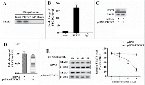
Figure 5. STAT3 promoted expression of INO80. (A) The chromatin immune-precipitation was used to verify the binding between PTCSC3 and the promoter of STAT3. (B) The expression levels of INO80 mRNA and protein in FTC238 cells were examined by qRT-PCR and western blot, respectively. **P < 0.01 vs. pcDNA. (C) The expression levels of INO80 mRNA and protein in 8505C cells were examined by qRT-PCR and western blot, respectively. **P < 0.01 vs. si-control.
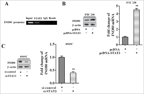
Figure 6. LncRNA PTCSC3 regulated INO80 through STAT3. (A) The expression levels of INO80 mRNA and protein in FTC238 cells were examined by qRT-PCR and western blot, respectively. **P < 0.01 vs. si-control. ##P < 0.01 vs. si-PTCSC3. (B) The expression levels of INO80 mRNA and protein in 8505C cells were examined by qRT-PCR and western blot, respectively. **P < 0.01 vs. pcDNA. ##P < 0.01 vs. pcDNA-PTCSC3.
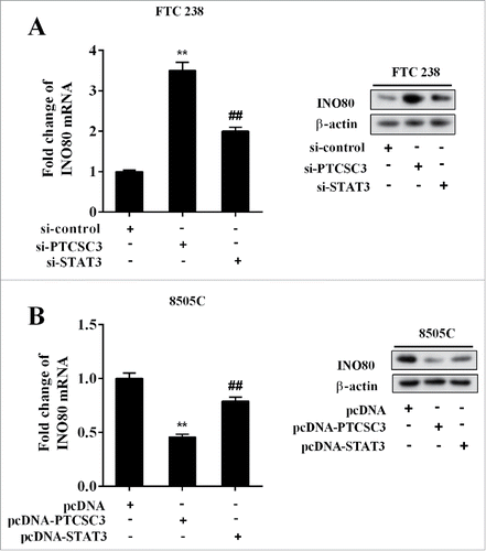
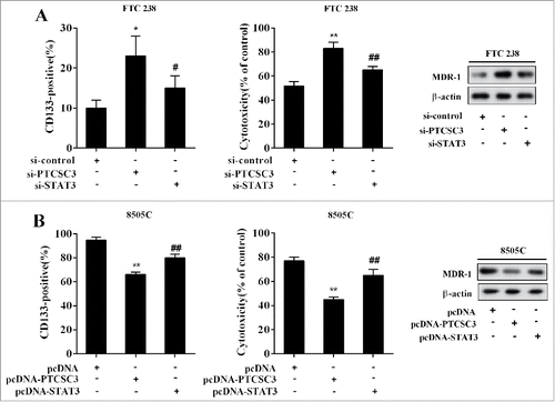
Figure 7. LncRNA PTCSC3 suppressed stem cells characteristics and drug resistance of ATC. (A) The positive expression rate of CD133 was analyzed by flow cytometry. *P < 0.05 vs. si-control; #P < 0.05 vs. si-PTCSC3; The cytotoxicity of DOX to FTC238 was detected by MTT assay. **P < 0.01 vs. si-control; ##P < 0.01 vs. si-PTCSC3; the levels of MDR-1 was analyzed by western blot. (B) The positive expression rate of CD133 was analyzed by flow cytometry. **P < 0.01 vs. pcDNA; ##P < 0.01 vs. pcDNA-PTCSC3; The cytotoxicity of DOX to FTC238 was detected by MTT assay. **P < 0.01 vs. pcDNA; ##P < 0.01 vs. pcDNA-PTCSC3; the levels of MDR-1 was analyzed by western blot.