Figures & data
Figure 1. CT scan before apatinib treatment (the lesions are indicated by arrows). A. CT scans before Apatinib therapy revealed disease progress in the Vaginal stump; B. after four cycles of gemcitabine, the metastatic mass became bigger; C. after three cycles of paclitaxel plus nedaplatin, the metastatic mass was still bigger; D. after oneone cycle of topotecan plus endostar, the metastatic mass continue to be bigger.
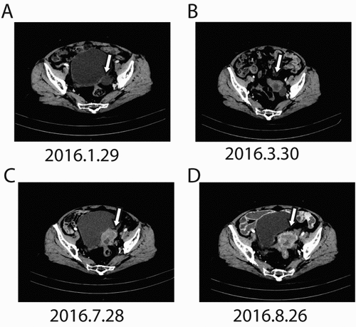
Figure 2. Changes in CA-125 levels before and after apatinib therapy. The CA-125 levels continuously increased from 28U/mL (Dec. 2015) to 2288 U/mL (Sep. 2016) before apatinib treatment. Apatinib plus epirubicin (solid arrows) began in Sep. 2016. Following initiation of this therapy, CA-125 levels dramatically decreased to 269 U/mL (Mar. 2017). Then, Apatinib monotherapy was used as maintenance therapy (open arrows), CA-125 levels come into a stable level.
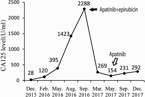
Figure 3. CT scan before apatinib treatment (the lesions are indicated by arrows). A. after five cycle of apatinib combined with epirubicin, CT scans showed that the metastatic mass became smaller significantly; B after more than four months of Apatinib monotherapy, CT scans showed that the metastatic mass in the vaginal stump was stable. C. after another about four months of Apatinib monotherapy, the mass in the vaginal stump was stable but the lymph node in front of the left external iliac artery increased. D. after another three months, bothe the lesion in the vaginal stump and front of the left external iliac artery were both stable.
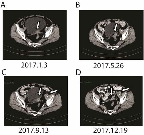
Table 1. The mutations detected by next-generation sequencing (NGS)
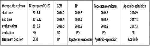
Figure 4. The various treatments the patient received as well as the duration of each treatment.