Figures & data
Figure 1. A tough mass measuring about 8 cm×9 cm was seen in the right eye, accompanied with tenderness and surface rupture.

Figure 2. Orbit computed tomography (CT) with intravenous contrast showed a 7.5-cm- × 4.1-cm-sized mass arising from the right orbit. The mass extended toward the right-sided maxillary sinus, ethmoid sinus, and frontal sinus.
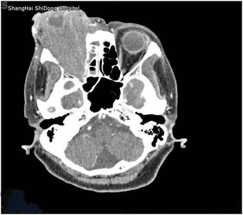
Figure 3. The tumor measuring 14 cm (a) × 15 cm (b) extended onto the right nasal cavity, which led to the shortness of breath.
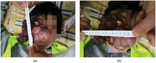
Figure 4. Contrast-enhanced CT imaging of the head revealed the tumor occupied the right orbit and was involved in the nasal cavity.
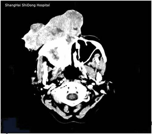
Figure 5. At the end of radiotherapy, the tumor was remarkably decreased in size.
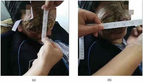
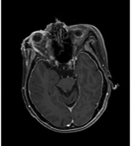
Figure 6. MRI image of the tumor prior to anlotinib hydrochloride. The tumor was located in the right orbit, with massive extension into the right maxillary sinus, ethmoid sinus, frontal sinus, sphenoid.
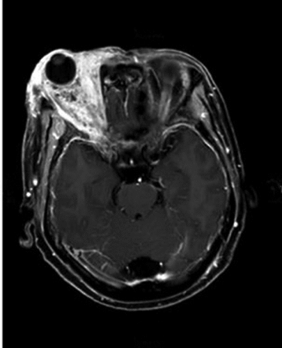
Figure 7. Two months later, the mass of the right orbit continued to shrink, with necrotic tissue on the surface.

Figure 8. Cranial MRI after four cycles of anlotinib hydrochloride, showing a significant reduction in the density of the right orbit malignance.