Figures & data
Figure 1. Up-regulated DLX6-AS1 promotes cell proliferation, migration and EMT in gastric cancer. (a) qRT-PCR analysis of the expression level of DLX6-AS1 in gastric cancer cell lines (AGS, HGC-27, BGC-823 and SGC-7901) and normal gastric epithelial cell GES-1. (b) DLX6-AS1 levels in AGS and HGC-27 cells with sh-DLX6-AS1#1, sh-DLX6-AS1#2 or sh-DLX6-AS1#3 treatment, compared with sh-NC group, were tested through qRT-PCR experiment. (c) CCK-8 assay was performed to detect the proliferative ability of DLX6-AS1-silenced AGS and HGC-27 cells. (d) Cell migration capacity of AGS and HGC-27 cells treated with sh-DLX6-AS1#1 was measured by transwell migration assay. (e) The expression levels of EMT-associated proteins, including E-cadherin, N-cadherin and Vimentin, in DLX6-AS1#1-transfected AGS and HGC-27 cells as respectively estimated by western blot. *P < .05, **P < .01.
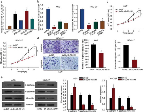
Figure 2. MAP4K1 highly expressed in GC is regulated by DLX6-AS1 in a positive way. (a) Western blot results of the expression of five MAP4K family members: MAP4K1, MAP4K2, MAP4K3, MAP4K4 and MAP4K5, under the transfection of sh-DLX6-AS1#1. (b) The expression level of MAP4K1 in GC cell lines and normal gastric epithelial cell was individually assessed by qRT-PCR. (c) qRT-PCR analysis of the levels of MAP4K1 in AGS and HGC-27 cells with the silencing of DLX6-AS1. *P < .05, **P < .01, ***P < .001.
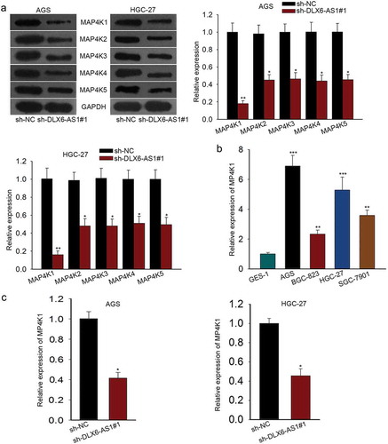
Figure 3. MAP4K1 enhances cell proliferation, migration and EMT in gastric cancer. (a) qRT-PCR result of MAP4K1 expression in AGS and HGC-27 cells with sh-MAP4K1#1/2/3 transfection. Here, sh-MAP4K1#1 transfection induced an obvious decrease in MAP4K1 expression. (b) CCK-8 experiment was carried out to evaluate the proliferation capacity of MAP4K1-silenced AGS and HGC-27 cells. (c) The migratory ability of AGS and HGC-27 cells with sh-MAP4K1#1 treatment was detected by transwell migration assay. (d) The levels of EMT-associated proteins, containing E-cadherin, N-cadherin and Vimentin, in sh-MAP4K1#1-transfected AGS and HGC-27 cells as tested by western blotting. *P < .05, **P < .01.
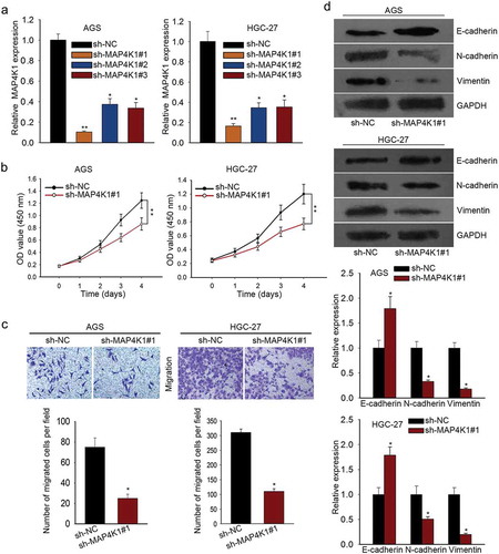
Figure 4. DLX6-AS1 stabilizes MAP4K1 by regulating FUS. (a) The binding of FUS and MAP4K1 from starbase v2.0. (b) RIP assay was conducted to confirm the combination of FUS with MAP4K1. (c) qRT-PCR analysis of the high FUS expression in GC cell lines. (d-e) The mRNA and protein levels of FUS in AGS and HGC-27 cells under sh-DLX6-AS1#1 treatment were evaluated by qRT-PCR. (f) AGS and HGC-27 cells were co-transfected with sh-DLX6-AS1#1 and pcDNA3.1 or sh-DLX6-AS1#1 and pcDNA3.1-FUS, with sh-NC and pcDNA3.1 as negative control. The efficiency of transfection was examined by qRT-PCR experiment. (g) Under actinomycin D treatment, the mRNA level of MAP4K1 with different treatments was dissected by qRT-PCR. *P < .05, **P < .01.
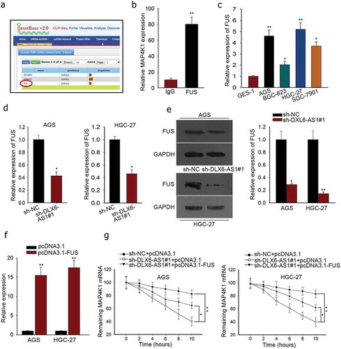
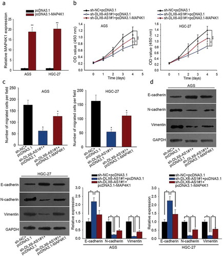
Figure 5. DLX6-AS1 improves cell proliferation, migration and EMT in GC by regulating MAP4K1. Cells were co-transfected with sh-DLX6-AS1#1 and pcDNA3.1 or sh-DLX6-AS1#1 and pcDNA3.1-MAP4K1, with sh-NC and pcDNA3.1 as normalization control. (a) MAP4K1 expression under pcDNA3.1-MAP4K1 transfection was confirmed by qRT-PCR. (b) CCK-8 experiment was applied to measure cell viability. (c) Cell migration was assessed by transwell migration assay. (d) Western blot analysis of EMT-associated proteins. *P < .05, **P < .01.