
Figures & data
Figure 1. Treatment timeline (Op: operation, RT: radiotherapy).
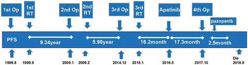
Figure 2. (a) The brown–yellow granules of the cell membrane and cytoplasm of vascular endothelial cells showed positive VEGFR2 (a × 100). Contrast-enhanced MRI (ceMRI) before treatment: transversal (b), and sagittal (c). ceMRI after treatment: transversal (e), and sagittal (f). e pathology of AE grade 3 (d × 200).
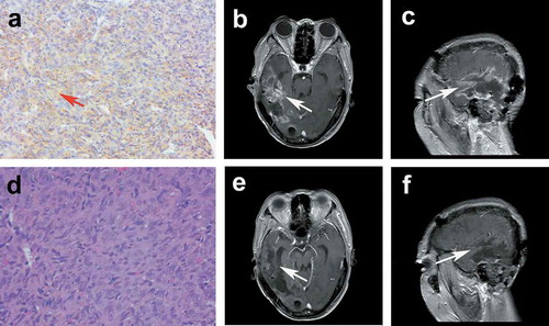
Figure 3. Treatment timeline (Op: operation; SRS: stereotactic radiosurgery).
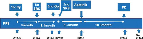
Figure 4. The brown–yellow granules of the cell membrane and cytoplasm of vascular endothelial cells showed a positive VEGFR2 (×200).
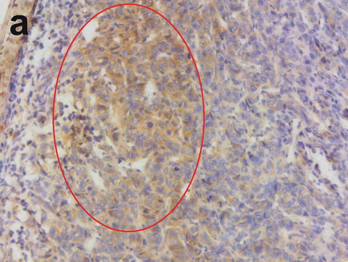
Figure 5. ceMRI before apatinib treatment: transversal (a), and sagittal (b). ceMRI after treatment: transversal (d), and sagittal (e). Flair images showed a significant relief of peritumoral edema (before treatment c vs. after treatment f).
Figure 6. Treatment timeline (Op: operation; IMRT: intensity-modulated radiation therapy).
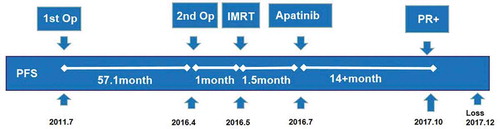
Figure 7. The brown–yellow granules of the cell membrane and cytoplasm of vascular endothelial cells showed a positive VEGFR2 (×200).
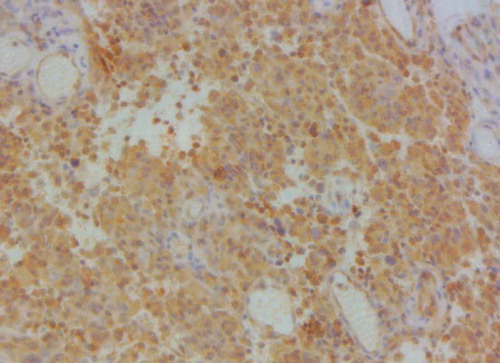
Figure 8. Postoperative ceMRI: transversal view (a), coronal view (b), and sagittal view (c). Postradiotherapy ceMRI: transversal view (d), coronal view (e), and sagittal view (f). ceMRI after treatment: transversal view (g), coronal view (h), and sagittal view (i).
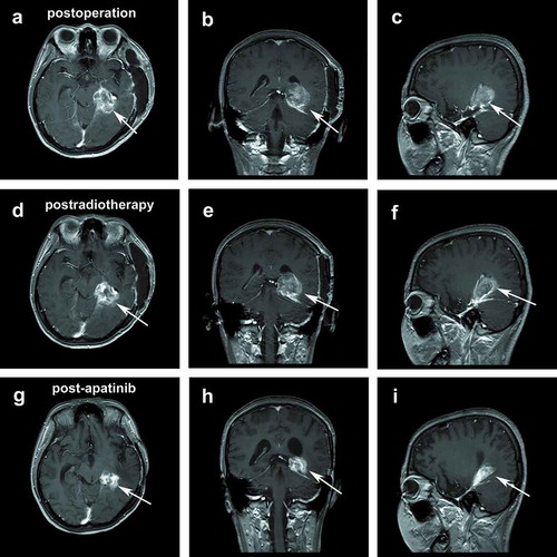
Table 1. Studies of VEGF/VEGFR inhibitors in treating refractory recurrent meningioma.
Availability of data and materials
The datasets used and/or analyzed during this study are available upon request from the corresponding author.